ATRIOVENTRICULAR SEPTAL
DEFECTS
 ATRIAL SEPTAL DEFECT
Definition, Spectrum of Disease, and Incidence
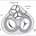
An atrial septal defect (ASD) is an opening of the atrial septum leading to a communication between the left and right atrium. According to its location, an ASD is classified into septum primum, septum secundum, sinus venosus, and coronary sinus defect (Fig. 15-1). Septum secundum defect (ASD II), which is due to lack of tissue in the region of the foramen ovale, is the most common, accounting for about 80% of all ASDs (1). Septum primum defect (ASD I), the second most common, represents a gap in the embryologic septum primum region, adjacent to both atrioventricular valves (Fig. 15-2) and is considered a partial atrioventricular septal defect (discussed later in this chapter). Sinus venosus atrial septal defect is found posterior and superior to the foramen ovale and inferior to the connections of the superior and inferior vena cava within the right atrium (Fig. 15-1). The rare coronary sinus atrial septal defect is located at the site of the coronary sinus ostium in the right atrium and is often associated with coronary sinus abnormalities (Fig. 15-1). The incidence of an ASD in postnatal series is high, accounting for 7% of all infants with congenital heart defects and occurring in 1 per 1500 live births with a 2:1 female-to-male ratio (2,3). ASD II and sinus venosus defects are practically inexistent in prenatal series (4). ASD I, detectable prenatally, is discussed in the section on atrioventricular septal defect.
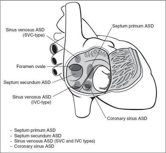
Figure 15-1. Types and anatomic locations of atrial septal defects (ASD) as seen from the right atrium. SVC, superior vena cava; IVC, inferior vena cava.
Stay updated, free articles. Join our Telegram channel

Full access? Get Clinical Tree


