Dental X-rays provide dentists with a clear view of the teeth, gums, and bone structure, allowing accurate diagnosis of oral health problems that aren’t visible during a routine examination.
X-rays are taken on an as-needed basis, reserved for new patients, those with advanced dental problems, oral surgery participants, and patients with good oral health on an annual or bi-annual basis. Dentists can take several different types of dental X-rays. In this blog, we’ll look at the types of radiology, when and how they are used, and their benefit in improving your oral health and beautiful smile, as told by dentist Dr. Broadbent.
Dental X-rays Detect Numerous Oral Health Issues
Dental X-rays allow your dentist to closely examine areas of your teeth, gums, and jaw bone that he cannot see with his eyes. Special X-rays are designed to show specific parts of the teeth, gums, or jaw and can help dentists detect numerous issues, including:
- Gum Disease: Also called periodontal disease, gum disease progresses through two sages. The first is gingivitis. When left untreated, gingivitis develops into gum disease. Dental X-rays help detect bone loss, tooth instability, and other symptoms of gum disease.
- Cavities: Acid is the primary cause of cavities. Sugar also contributes. Most form under the gum line or between the teeth, which dentists cannot see during a visual exam.
- Tooth Position: Dental X-rays help dentists track tooth development and tooth position, helping better identify misalignment and similar issues.
- Infections: Infections and abscesses within the bone or jaw structure cannot be detected through visual inspection. An X-ray helps make an accurate diagnosis.
Dental X-rays allow early detection and treatment of issues listed above and many others. Patients can smile more easily knowing dental care is on the way.
Types of Dental X-rays
There are numerous types of dental X-rays. Each type serves a unique purpose. Let’s look at the types of X-rays and learn more about how dentists use them.
- Bitewing X-ray
The Bitewing X-ray is the most commonly used type of dental X-ray. It is used during routine dental checkups, including for first-time patients. This X-ray provides an image of the upper and lower back teeth on one film and images of the spaces between the teeth.
The Bitewing X-ray helps dentists spot cavities between the teeth, including the back molars. Bitewing X-rays also allow dentists to evaluate the health of the bone structure supporting the teeth, detect bone loss, and identify decay.
Quick and easy to perform, the bitewing X-ray helps dentists spot tooth decay and other issues in seconds. Dentists need to help provide patients with maximum oral health protection.
- Periapical X-rays
Another type of dental X-ray is the Periapical type. This X-ray takes a close-up image of an entire tooth, its root, and surrounding bone. It gives dentists a focused view of an area, allowing them to diagnose issues with the tooth root or surrounding area. It helps diagnose abscesses, tumors, bone loss, cysts, and similar structural issues.
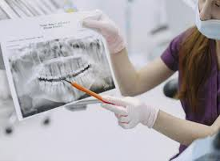
- Panoramic X-ray
A panoramic X-ray takes an image of a patient’s entire mouth, including the sinuses and nasal area. This X-ray benefits a patient who needs braces or dental implants or has tumors or jaw issues. It can also benefit dentists when evaluating wisdom teeth and help assess alignment issues.
Unlike other dental X-rays, this one captures the entire mouth in one image. Dentists can use the image to diagnose the overall oral health of the patient’s mouth.
- Occlusal X-rays
A fourth type of dental X-ray is the Occlusal X-ray. This X-ray examines the bite of a patient or the position of the teeth related to one another. It allows dentists to diagnose more serious problems like cysts or jawbone-related issues.
The Occlusal X-ray creates an image of the patient’s upper or lower jaw and shows the teeth alignment and the condition of the bone structure.
- CBCT Scans
A Cone Beam CT scan, commonly known as a CBCT can, is designed for complex oral health issues, including jaw surgery, root canals, and dental implants. It takes a 3D image of a patient’s mouth when dentists require high-precision imaging with even greater detail than traditional X-ray imagery. It can provide insight into the soft tissues, nerve pathways, and bone structure of the mouth.
Which X-ray is Best For Your Dental Situation?
Your dentist will determine which type of X-ray is best for your situation based on the reason for your visit and your current oral health condition. Most patients take the bitewing X-ray; it is common during regular exams.
Dental X-rays FAQ
- Are dental X-rays safe? Yes, dental X-rays are safe when completed by a trained professional. Although X-rays use radiation, it’s in amounts so small there is no risk to humans. Dentists take additional protective measures to safeguard patients and their teams, further reducing any potential risks associated with the procedure. Rest assured, dental X-rays are completely safe.
- Does insurance cover all types of X-rays? Dental insurance covers traditional routine X-rays like the Bitewing X-ray, but may not cover more advanced imaging, such as the CBCT scan. Check your insurance policy or speak to a representative to determine what your policy covers.
- How long does it take to complete a dental X-ray? You’ll sit in the office waiting for the dentist longer than it takes to complete an X-ray. The common X-ray, the Bitewing, takes an average of 5 to 10 minutes from start to finish, and the others are similarly quick and easy.
- Are dental X-rays painful? Dental X-rays are not painful. It is nothing more than taking a photograph of your mouth!
The Bottom Line
Dental X-rays give dentists extra help in protecting your oral health. Each type serves a unique purpose, but rest assured, it benefits your smile tenfold. We hope this blog provides information about dental X-rays that eases your mind and gives you a clearer understanding of their importance.
Related posts:


Stay updated, free articles. Join our Telegram channel

Full access? Get Clinical Tree


