9 White-out hemithorax
Background
They can be differentiated by clinical and radiological features, and by complex imaging.
Radiological features
The key to interpretation is the position of the mediastinum.
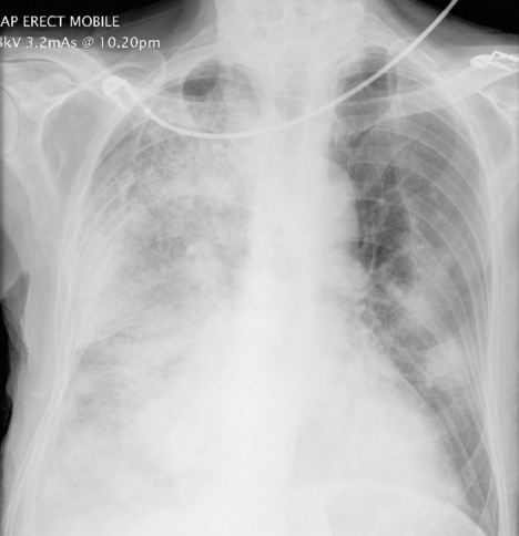
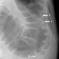
In pneumonia (Fig. 9.1), there is no loss of volume in the lung so the mediastinum and trachea remain central. The other clue in pneumonia is that the shadowing may be a little patchy and there may be air bronchograms.
Stay updated, free articles. Join our Telegram channel

Full access? Get Clinical Tree


