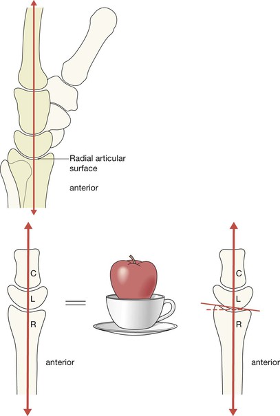
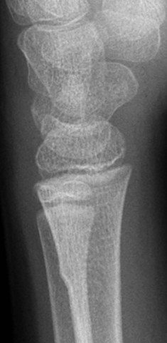
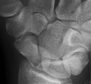
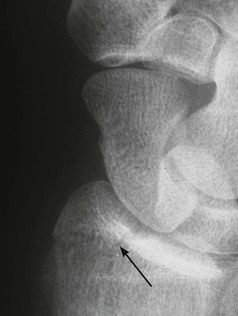
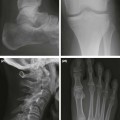
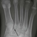
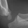
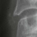
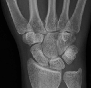
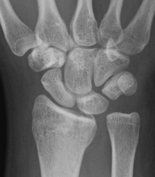
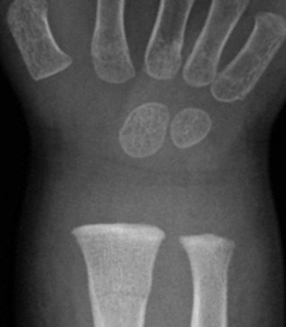
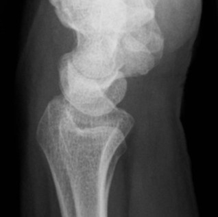
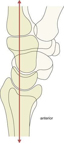
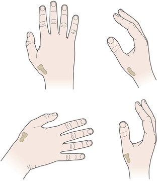
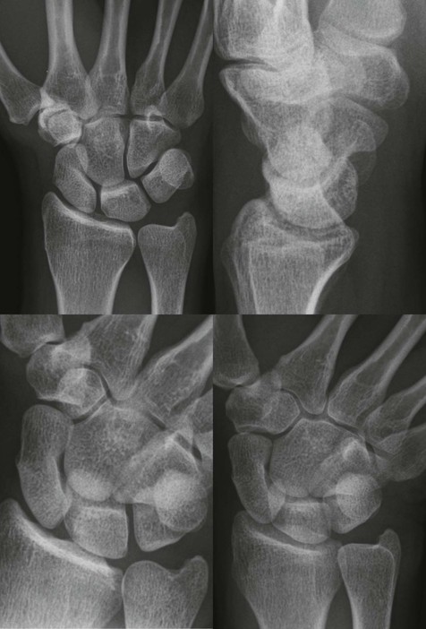
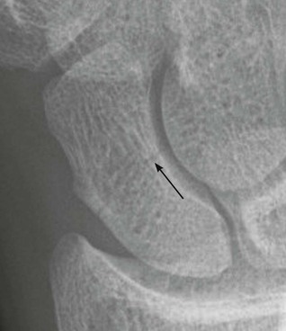
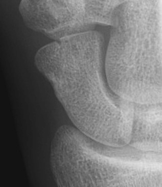
The articular surface of the radius lies distal to that of the ulna in 90% of normal people. The carpal bones are arranged in two rows, bound together by strong ligaments: ▪ The joint spaces are uniform in width: 1–2 mm wide in the adult. ▪ Adjacent bones have parallel/congruous surfaces. ▪ Abnormally wide spaces are likely to indicate damaged ligaments. The dorsal cortex of the distal radius is completely smooth—no crinkles, no irregularity. This cortex should be as smooth as a baby’s bottom. The alignment of the carpal bones may appear confusing but identifying the important anatomy is actually very simple. Don’t worry about the overlapping bones. Just think: apple, cup, saucer. The PA view will appear fairly comforting to an inexperienced observer because all of the carpal bones are clearly shown. The lateral radiograph may appear terrifyingly complex and difficult to analyse because of the numerous overlapping bones. There is a very clear message: do not be afraid! The lateral view is diagnostically very, very, important, so we will show you how to quickly and confidently analyse every lateral radiograph using a simple checklist. Analysis: ask yourself five questions. Questions 1–4 apply to all adults. Question 5 applies to all children. 1. Is the radial articular surface and/or the ulna styloid process whole and intact? 2. Does the radial articular surface lie distal to the ulna? 3. Is the scaphoid bone intact and normal? 4. Is the scapho-lunate distance less than 2 mm wide? 5. In children: does the radial cortex show any angulation or any suggestion of a localised bulge? Normal PA wrist. The answer to questions 1–4 is yes. Abnormal PA wrist. (1) Subtle increase in density of the metaphysis of the radius suggests an impacted fracture. (2) Widening of the distal radio-ulnar joint and the ulnar articular surface lies distal to the immediately adjacent radial articular surface. The radio-ulnar joint is disrupted. Abnormal PA wrist. A subtle lucent line crosses the metaphyseal–diaphyseal region of the radius. Note the slight bulging of the adjacent cortex. Fracture of the radius. A Torus fracture (p. 18) Analysis: ask yourself five simple questions on each and every lateral view. No exceptions. If you ask and correctly answer these five questions you will detect all the subtle and clinically important abnormalities. 1. Is the radial articular surface intact? 2. Is the dorsal cortex of the distal radius smooth? Specifically: □ Is the cortex as smooth as a baby’s bottom? □ No crinkle, no angulation, no bulge, no buckling? □ Are you sure? Check the dorsal cortex one more time. 3. Is the palmar tilt (normal range 2–20°) of the articular surface of the radius normal? 4. Is there a bone fragment lying posterior to the carpal bones? 5. Is there a bone sitting in the cup of the lunate? Normal lateral wrist. Normal palmar tilt. Dorsal cortex of the radius is as smooth as a baby’s bottom. The cup of the lunate is full—it articulates normally with the capitate. The three bones (radius, lunate, capitate) are in line. Normal appearance. Many undisplaced scaphoid fractures are not visualised on the two standard (wrist) views. Two extra views produces a better return. Therefore, a four view scaphoid series is essential and should be requested whenever there is ‘snuffbox’ tenderness: The two additional images will vary between Emergency Departments. Importantly, two of the four projections will always include a true PA and a true lateral of the wrist. Scaphoid fractures are mainly hairline fractures and lucent; they are not sclerotic. Occasionally the fracture is displaced. Analysis: ask yourself three questions. 1. Does the scaphoid appear intact on each of the four views? 2. Is the distal radius—particularly the styloid process—intact? 3. Have I checked the PA and lateral views step-by-step (see pp. 128–130)? Snuffbox tenderness. A four view scaphoid series. A four view scaphoid series. Normal. Hairline fracture (arrow) through the waist of the scaphoid. This is the most typical appearance. Fracture of the distal pole of the scaphoid. Distal pole fractures are comparatively infrequent.
Wrist & distal forearm
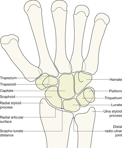
Normal anatomy
PA projection: bones and joints
Lateral projection: bones and joints
Analysis: the checklists
The PA view
No = undisplaced fracture.
No = suspect disruption at the radio-ulnar joint.
No = fracture.
No = suspect a tear of the scapho-lunate ligaments (p. 147).
Yes = Greenstick or Torus fracture.
The lateral view
No = undisplaced fracture.
No, it is not smooth = undisplaced fracture.
No = suspect an impacted fracture.
Yes = Triquetral fracture.
No = carpal dislocation involving the lunate (pp. 148–150).
The scaphoid series
No = fracture (see p. 144).
No = fracture (see pp. 136–141).
AND
No = start checking.

Wrist & distal forearm
9