ZMC Fracture
Kwaku A. Obeng
CLINICAL HISTORY
29-year-old male involved in a motor vehicle accident. The patient does not recall striking his head, but complains of tenderness over the right zygomatic arch and maxilla.
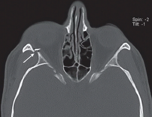 FIGURE 21A |
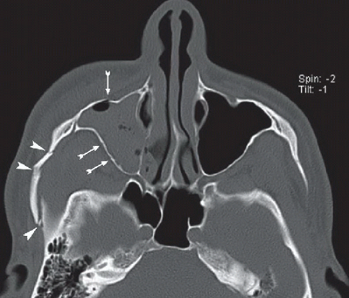 FIGURE 21B |
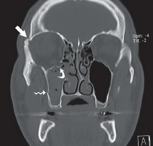 FIGURE 21C |
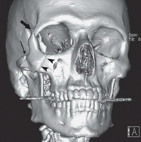 FIGURE 21D |
FINDINGS
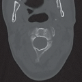
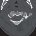
Figures 21A and 21B: Axial noncontrast maxillofacial CT images. Figure 21A demonstrates a comminuted fracture (thin white arrow) through the right lateral orbital wall involving the sphenozygomatic suture, with posterolateral displacement of the lateral orbital rim, and a medially displaced and laterally angulated comminution fragment encroaching on the orbit. In Figure 21B, there is a comminuted fracture of the right zygomatic arch (white arrowheads), including a more posterior fracture through the base of the zygomatic process of the temporal bone. There are also nondisplaced fractures through the anterior and posterolateral walls of the right maxillary sinus (hatched arrows). Figure 21C: Coronal reformatted CT image demonstrates an additional fracture of the right superolateral orbital rim (thick white arrow), just above the frontozygomatic suture. There is a comminuted, inferiorly displaced fracture through the right
orbital floor (curved arrow) involving the infraorbital nerve canal, and a minimally displaced fracture of the lateral wall of the right maxillary sinus (wavy arrow). Figure 21D: Right anterior oblique projection of a 3D surface shaded rendering demonstrates fractures of the right lateral (thick black arrow) and inferior orbital rims (black arrowheads) and right zygomatic arch (thin black arrow).
orbital floor (curved arrow) involving the infraorbital nerve canal, and a minimally displaced fracture of the lateral wall of the right maxillary sinus (wavy arrow). Figure 21D: Right anterior oblique projection of a 3D surface shaded rendering demonstrates fractures of the right lateral (thick black arrow) and inferior orbital rims (black arrowheads) and right zygomatic arch (thin black arrow).
Related posts:


Stay updated, free articles. Join our Telegram channel

Full access? Get Clinical Tree


