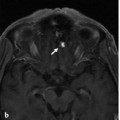
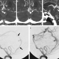
The Azygos Anterior Cerebral Artery A 54-year-old woman was investigated for recurrent migraines. Unenhanced computed tomography at an outside institution had revealed an aneurysm. See ▶ Fig. 12.1. Fig. 12.1 3D CTA reconstructions (a,b) demonstrate an azygos ACA (arrowheads) with a distal aneurysm in the callosomarginal artery bifurcation (arrows). Azygos anterior cerebral artery (ACA) with a callosomarginal bifurcation aneurysm. The azygos ACA is a relatively uncommon vascular anomaly, with a prevalence ranging between 0.2% and 4%. Embryologically, the azygos vessel appears to form via fusion of the A2 segment, which originates either from the medial branch of the olfactory artery at the 16-mm stage of embryogenesis (40 days of gestation) or from the continuation of the median artery in the corpus callosum at the 20–24-mm stage. According to the Baptista classification, the azygos ACA is type 1 when there is a single unpaired ACA, as in the present case description. In type 2, there are bihemispheric ACAs, one of which is dominant and supplies both hemispheres. Finally, in type 3, there is an accessory ACA in the presence of a median artery that arises from the anterior communicating artery. Type 2 has an estimated prevalence of 2 to 7%. It can be differentiated from the azygos ACA by the presence of a hypoplastic A2 segment. Its main clinical relevance is that occlusion of the dominant A2 segment will result in bilateral ACA territory infarcts. Type 3 has also been termed ACA trifurcation. The ACA trifurcation is defined by three A2 segments arising from the anterior communicating artery. It has an estimated prevalence of 2 to 13%. This normal variant may be explained by persistence and/or enlargement of the median callosal artery (▶ Fig. 12.2). Fig. 12.2 A 76-year-old woman presented with left watershed territory strokes. CTA 3D volume reconstruction in anteroposterior (AP) view shows three branches coming out of the anterior communicating artery complex region, in keeping with an ACA “trifurcation.”
12.1 Case Description
12.1.1 Clinical Presentation
12.1.2 Radiographic Studies

12.1.3 Diagnosis
12.2 Embryology and Anatomy

Related posts:


Stay updated, free articles. Join our Telegram channel

Full access? Get Clinical Tree


