The insertions of the supraspinatus and infraspinatus tendons have been further described as a five-layer structure (table 1-2).4 Layer two comprises the main portion of the tendons, though layer four deserves special consideration because of its crucial functional role (figure 1-1). The layer four is consistently located at the margin of the critical zone (see section 2.2), extends from its anterior attachment just posterior to the biceps tendon to its posterior attachment near the inferior border of the infraspinatus tendon,5 and function in a way that is analogous to a load-bearing suspension bridge, distributing load and stress-shielding the distal hypovascular tendon.6 Hence, ruptured supraspinatus tendon can still exert most of its functions as long as layer four is intact and tear restricted to the distal fibers. This mechanism may limit the propagation of distal tears as well as explain why some tears have preserved biomechanics (figure 1-2).
Table 1-2. The five histological layers of the supra- and infraspinatus tendons, from superficial to deep.
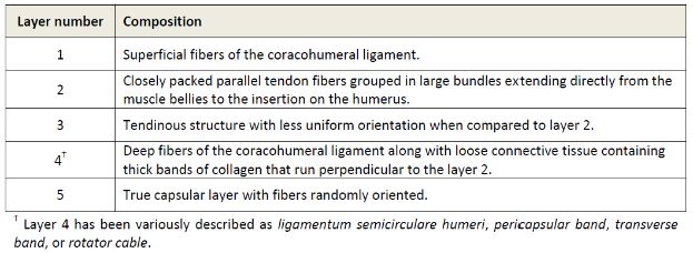
Figure 1-1. Shoulder abduction. Schematic drawing of an axial view demonstrates shoulder abduction [A] in the coronal and [B] scapular plane. The scapular plane is anteriorly rotated about 30o with respect to the coronal plane. Adduction or abduction of the shoulder in the scapular plane is also known as scaption.
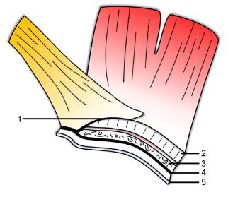
Figure 1-2. Five-layer structure of rotator cuff. Schematic drawing demonstrates the five-layer structure of the insertion of the supraspinatus and infraspinatus tendons. See table 1-2 for details.
As a whole, the rotator cuff muscles perform multiple functions, including abduction, internal rotation, and external rotation of the shoulder. The rotator cuff also stabilizes the glenohumeral joint and controls humeral head translation. The infraspinatus and subscapularis have significant roles in scapular plane shoulder abduction, generating forces that are two to three times greater than the force produced by the supraspinatus muscle (figure 1-3).7 However, the supraspinatus is more effective for general shoulder abduction because of its moment arm. The anterior portion of the supraspinatus tendon is submitted to significantly greater load and stress, and performs its main functional role.8 The rotator cuff is responsible for up to 50% of muscle effort for shoulder abduction and 80% for external rotation.9
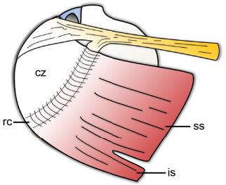
Figure 1-3. Rotator cable. Schematic drawing of a superior view of the shoulder. The rotator cable (rc) is located at the margin of the critical zone (cz) and extends from its anterior attachment just posterior to the biceps tendon to its posterior attachment near the inferior border of the infraspinatus tendon (is). Ss= supraspinatus. Yellow= coracohumeral ligament.
Related posts:

Stay updated, free articles. Join our Telegram channel

Full access? Get Clinical Tree


