Discussion
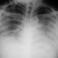
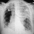
This patient’s film reveals an extensive confluent infiltrate in the right lower lung field. One could assume that this represents an extensive pneumonia. There are however, are other patchy infiltrates, including one in the left upper lobe and probably an infiltrate of the left lower lobe, posteriorly, behind the heart. Note that there are air bronchograms in the right lower lobe. The patient was not running a fever, and his white count was within normal limits. The findings most likely represent ARDS.
This 32-year-old patient had a cholycystectomy, subsequently developed a bile leak, and had to go back to the OR. Progressive respiratory problems ensued, and this film was obtained 5 days later.
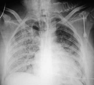
Discussion
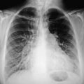
The film reveals extensive confluent bilateral infiltrates. Do not mistake this for failure. The heart is normal, and the infiltrates are not perihilar. They are more extensive in the right lower lobe, with several patchy areas in the right upper lobe, and extensive alveolar changes on the left. Previously, the patient had several small areas of infiltrate, but now there is coalescence and extension. This is all due to an alveolar leak and extensive ARDS. A left-sided central line, placed via the internal jugular route, is seen. The endotracheal tube is in fairly good position, but probably slightly higher than one would like to see.
This 62-year-old patient with pancreatitis was admitted to the hospital and developed severe respiratory difficulty after 7 days.