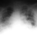
Discussion
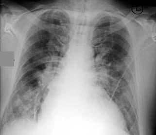
Portable film reveals the heart to be at the upper limits of normal. Note the classic appearance of pulmonary edema with bilateral perihilar fluid, loss of definition of the pulmonary vascularity, and a fairly classic bat wing appearance. The patient has had an intraortic pump balloon inserted, which is in good position. A central line and endotracheal tube are also in good position.
This patient, who has severe coronary artery disease and congestive myopathy, was admitted to the ICU.
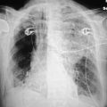
Discussion
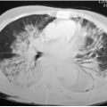
This patient has an enlarged heart and fairly classic findings of increased flow to the upper lobe, so-called cephalization, as well as perivascular cuffing.
This patient entered the emergency department with dyspnea.
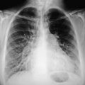
Discussion
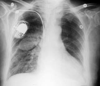
The patient has a bipolar pacer. What is interesting is the density in the right middle lung field. This elliptical density represents fluid trapped the oblique fissure. Normally fluid is free in the pleural space, but here fluid accumulates between the two layers of the pleura and is “pinched” off in two areas due to fibrosis and scarring, so a pool of fluid develops in this area, producing this characteristic appearance. This may disappear spontaneously and rapidly, and this density is sometimes referred to as a vanishing tumor.
Related posts:

Stay updated, free articles. Join our Telegram channel

Full access? Get Clinical Tree


