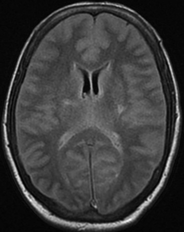
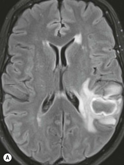
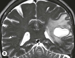
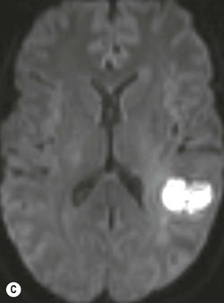
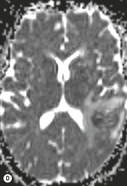
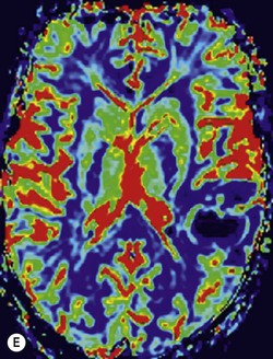
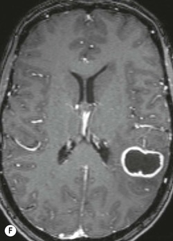
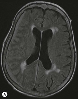
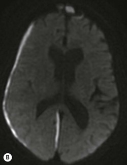
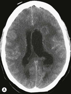
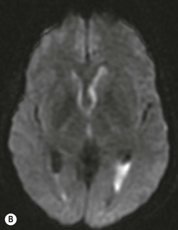
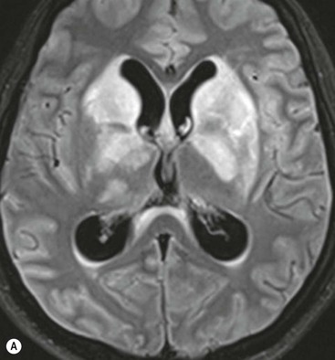
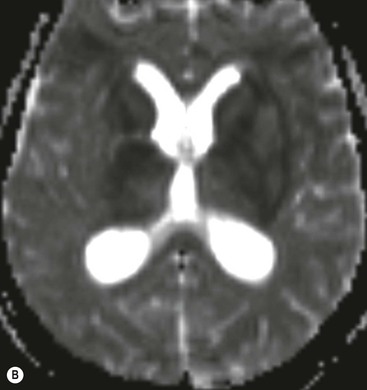
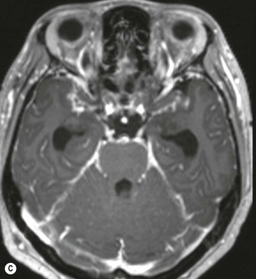
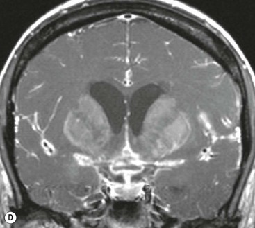
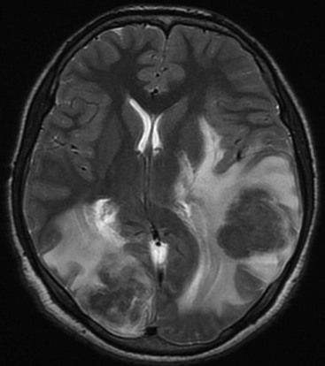
Daniel J. Scoffings, Julia Frühwald-Pallamar, Majda M. Thurnher, H. Rolf Jäger The causes of bacterial meningitis are age-dependent; in adults the most common causes are Streptococcus pneumoniae and Neisseria meningitidis. Bacteria can reach the meninges by haematogenous dissemination, spread from an adjacent focus of infection (such as sinusitis or otomastoiditis) or through congenital or acquired structural defects in the skull. Clinical manifestations include fever, headache, photophobia, lethargy and confusion. Imaging is frequently normal in patients with uncomplicated bacterial meningitis and is not necessary for its diagnosis, which requires analysis of a sample of cerebrospinal fluid (CSF) obtained by lumbar puncture (LP). The issue of whether neuroimaging is necessary before LP can be a matter of dispute between referring clinicians and radiologists. Although computed tomography (CT) can show findings that contraindicate LP, such as effacement of the basal cisterns and cerebellar tonsillar herniation, a normal CT does not imply that LP can be performed without the risk of causing tonsillar herniation.1 The guidelines of the National Institute for Health and Clinical Excellence (NICE) in the United Kingdom are that CT is indicated when there are focal neurological signs or with a reduced or fluctuating level of consciousness, but that treatment should not be delayed in order to obtain imaging.2 Imaging is often normal in patients with uncomplicated bacterial meningitis but some abnormalities may be observed. Subtle distension of the subarachnoid spaces by inflammatory exudate has been reported, but can be difficult to appreciate in the very young and in older subjects, both of whom have relatively prominent basal cisterns. Leptomeningeal contrast enhancement, which follows the surface of the brain and extends into the sulci and basal cisterns, may be seen and is better detected by contrast-enhanced magnetic resonance imaging (MRI) than by contrast-enhanced CT. Leptomeningeal enhancement can be difficult to distinguish from contrast enhancement in normal blood vessels and the detection of abnormal enhancement of the leptomeninges may be improved by the use of a post-contrast fluid-attenuated inversion recovery (FLAIR) sequence, which is less sensitive to vascular enhancement.3 An unenhanced FLAIR sequence may also show increased signal intensity in the subarachnoid spaces, most often over the frontal convexities and in the sylvian fissures, as a result of increased protein concentration in the CSF, causing failure of signal suppression by the inversion pulse (Fig. 63-1).4 This FLAIR hyperintensity is non-specific, however, and can also be seen with subarachnoid haemorrhage, with malignant meningitis and in patients receiving large doses of supplemental oxygen. In a minority of cases (probably less than 10%) diffusion-weighted imaging (DWI) can show multiple foci of high signal intensity in the subarachnoid spaces; these are typically nodular and are associated with increased signal intensity on the FLAIR sequences in most cases. The finding of subarachnoid high signal on DWI has been reported to be associated with a poor prognosis.5 Complications of bacterial meningitis include hydrocephalus, most often of the communicating type, brain abscess, subdural empyema and ventriculitis (see below). Focal parenchymal lesions may also be observed on DWI, principally secondary to vasculopathy of arteries surrounded by inflammatory exudate, and have been classified into four patterns: large vessel territory infarcts, perforator territory infarcts, multiple bilateral lesions in the cortex and subcortical white matter and multiple bilateral lesions restricted to the cerebral cortex.6 In immunocompetent patients most brain abscesses are bacterial, streptococci accounting for the majority. In 20–40% no causative organism is identified. Brain abscesses arise by haematogenous dissemination, penetrating trauma or direct spread from contiguous infection. The site of an abscess depends on its cause: frontal sinusitis will result in an abscess in or beneath the adjacent frontal lobe, whereas mastoiditis will give rise to a temporal lobe or cerebellar lesion. Blood-borne infection can occur anywhere in the brain, but has a predilection for the territory of the middle cerebral arteries, particularly the frontoparietal region. A thorough search for a predisposing factor should be made; a cardiac cause is frequently overlooked (occult endocarditis and septal defects). Abscesses are frequently subcortical or periventricular. Four stages of development are described: early and late cerebritis and early and late capsule formation. Patients present with fever (in 50%), headache and focal neurological deficits. Brain abscesses are multiple in 10–50%.7 On CT, cerebritis appears as ill-defined low attenuation; enhancement is usually absent at the early stage but can appear irregular and peripheral in late cerebritis and may progress centrally on delayed images. With capsule formation the abscess shows central low attenuation, because of pus or necrotic debris and a rim of slightly higher attenuation surrounded by low-attenuation vasogenic oedema. After contrast medium, a ring of enhancement corresponds to the capsule. The enhancing rim typically has a smooth inner margin and shows thinning of its medial aspect.8 In contrast to cerebritis, the centre of the abscess never enhances on delayed images. The degree of enhancement is diminished in patients who are immunocompromised or are on corticosteroid therapy.9 Abscesses rarely contain gas; when present this is most often caused by surgical intervention or communication with a cranial air space such as the paranasal sinuses or mastoid air cells. It is only rarely because of a gas-forming organism. On MRI, the signal of the abscess centre is intermediate between that of CSF and white matter on T1-weighted images and iso- or slightly hyperintense to CSF on T2-weighted images. On T2 sequences the abscess rim is relatively hypointense (Fig. 63-2); it may be slightly hyperintense to white matter on T1 images.10 The abscess rim often appears markedly hypointense on susceptibility-weighted imaging (SWI); this is thought to be the result of free radicals produced by phagocytosis.11 A ‘dual rim’ sign of concentric outer hypointensity and inner hyperintensity relative to the abscess core can also be seen on SWI.12 The pattern of rim enhancement is similar to that shown by CT, the outer margin rim more often being smooth than lobulated.13 Surrounding vasogenic oedema is of low signal on T1 and high signal on T2 images. The abscess centre is of high signal on DWI and low signal on maps of apparent diffusion coefficient (ADC), because of restricted diffusion in the viscous pus (Fig. 63-2). The degree of restriction of diffusion is inversely correlated with the viable cell count within the abscess centre.14 Though typical, the appearance of a brain abscess as a rim-enhancing mass is non-specific and may be mimicked by metastasis, glioblastoma and resolving haematoma. A thick, irregular rind of enhancement is more suggestive of tumour. Abscesses are more likely to show small satellite lesions. Despite initial hopes that restricted diffusion would reliably distinguish abscess from tumour, reduced ADC has been subsequently reported in metastases and glioblastomas. Dynamic contrast-enhanced perfusion MRI may help distinguish between brain abscess and tumour; abscesses have a lower relative cerebral blood volume in their enhancing rim than gliomas.15 The management of brain abscesses can require both medical and neurosurgical therapy. CT diagnosis has been responsible for a marked reduction in the mortality of brain abscesses. Follow-up imaging is recommended at biweekly intervals or when new symptoms arise. Sufficient treatment is indicated by resolution of rim enhancement or disappearance of the low signal rim on T2 images. Treatment response may be better assessed with DWI than conventional MRI; low signal on DWI correlates with a good clinical response, whereas increasing signal implies reaccumulation of pus.16 An intracranial epidural abscess is a collection of pus between the inner table of the skull and the endosteum of the skull, which forms the outer layer of the dura mater. Abscesses mainly arise by direct spread from a contiguous focus of infection, most often sinusitis or otomastoiditis, less often as a complication of dental sepsis or after craniotomy. Streptococcus milleri is the most frequent causative organism. As intracranial epidural abscesses are typically slow-growing, the clinical presentation is often insidious, usually with fever and headache. CT and MRI show a lentiform collection of fluid constrained by the dura at the sites of cranial sutures. Accordingly, when anteriorly located as a complication of frontal sinusitis they can cross the midline, in contrast to subdural empyemas which do not cross the midline. The fluid within the epidural abscess is of slightly higher attenuation than CSF on CT and is hyperintense on T2 and FLAIR images and is slightly hyperintense compared to CSF on T1.17 The dura at the deep margin of an epidural abscess typically shows thick and slightly irregular contrast enhancement (Fig. 63-3). Similar to cerebral abscesses, the pus within an epidural abscess can show restricted diffusion, appearing hyperintense on DWI and hypointense on ADC maps.18 In patients with fever after a neurosurgical operation, normal and expected sterile fluid collections in the epidural space can be hard to distinguish from an epidural abscess, but a serial increase in the attenuation of the fluid suggests infection. DWI has been found to be less sensitive and specific in the postoperative setting.19 A subdural empyema is a collection of pus in the potential space between the inner layer of the dura mater and the arachnoid mater. Empyemas occur more commonly than epidural abscesses. As with epidural abscesses, the most frequent predisposing causes are sinusitis and otogenic infection; head trauma, surgery and haematogenous spread are less common. Headache, fever, focal neurological deficit and meningism are the most frequent clinical features at presentation. The CT and MRI appearances of subdural empyema are of a crescentic fluid collection overlying the cerebral convexity or in the interhemispheric fissure alongside the falx cerebri. The margins of the collection may be irregular and scalloped, as a result of loculation. Although contrast enhancement at the deep margin of a subdural empyema is a characteristic finding, it can be subtle or absent in the early stages of infection. The adjacent brain may show oedema or cortical contrast enhancement. The CT attenuation and MRI signal characteristics of a subdural empyema are the same as for an epidural abscess (Fig. 63-4). Ventriculitis is uncommon. Causes include trauma, intraventricular rupture of an abscess, shunt infection and haematogenous spread of infection to the ependyma or choroid plexus. The most frequent imaging finding is intraventricular debris, which is slightly hyperattenuating compared to CSF on CT and is of increased signal on FLAIR and DWI sequences with low signal intensity on ADC maps (Fig. 63-5). Periventricular and subependymal high signal and enhancement of the ventricular margins are less common although are still present in most cases.20,21 The affected ventricles are usually dilated. Involvement of the central nervous system (CNS) occurs in 5% of cases of tuberculosis; most patients are younger than 20 years. Of patients with CNS tuberculosis, the chest radiograph is abnormal in 45–60%.7 Tuberculous meningitis is the most frequent manifestation of tuberculous CNS infection. In the early stages of the disease a diffuse pattern of leptomeningeal enhancement is common, with a later predilection for the basal leptomeninges, most frequently in the interpeduncular cistern of the midbrain.22 CT shows obliteration of the basal cisterns by isoattenuating or slightly hyperattenuating exudate, which enhances diffusely after IV contrast medium. The most sensitive and specific CT criteria for tuberculous meningitis are linear enhancement of the middle cerebral artery cisterns, obliteration by contrast of the CSF spaces around normal vascular enhancement, Y-shaped enhancement at the junction of the suprasellar and middle cerebral artery cisterns and asymmetry of enhancement.23 The meningeal exudate obstructs the resorption of CSF and so causes communicating hydrocephalus, resulting in dilatation of the lateral, third and fourth ventricles. This is seen in 50% of adults and 85% of children.7 Less often, non-communicating hydrocephalus can occur because of obstruction of the outlet foramina of the fourth ventricle. Arteritis of the penetrating arteries within the subarachnoid space affected by areas of tuberculous meningitis can result in infarctions of the basal ganglia, internal capsules and brainstem. With healing, calcification of the affected meninges may be seen rarely. MRI depicts the basal meningeal enhancement, hydrocephalus and perforator territory infarcts with greater sensitivity than CT (Fig. 63-6). The differential diagnosis for tuberculous meningitis includes fungal meningitis, neurosarcoid and carcinomatous meningitis. Tuberculomas (parenchymal granulomas) occur most often at the junction of white and grey matter (Fig. 63-7). On CT they appear as small rounded lesions isoattenuating or hypoattenuating to normal brain, with variable amounts of surrounding vasogenic oedema. Contrast enhancement is homogeneous when lesions are solid and shows rim enhancement when central caseation or liquefaction occurs. The ‘target sign’ of central high attenuation with rim enhancement is not pathognomonic for tuberculoma. On MRI small, non-caseated, tuberculomas show low signal intensity on T1 sequences and high signal on T2 sequences. Caseation of tuberculomas results in low signal intensity on T2 (Fig. 63-7). On DWI tuberculomas may show elevated or restricted diffusion. Tuberculomas may calcify when healed, but, as with meningeal disease, this is uncommon.24 Tuberculous abscesses are uncommon; they may resemble tuberculomas but are usually larger and have a thinner enhancing rim. The enhancing rim of a tuberculous abscess is more often lobulated than smooth, whereas, similar to pyogenic abscesses, the non-enhancing core of a tuberculous abscess typically shows restricted diffusion.13
Intracranial Infections
Bacterial Infections
Bacterial Meningitis
Cerebritis and Brain Abscess
Epidural Abscess and Subdural Empyema
Ventriculitis
Tuberculosis
Neurosyphilis
Related posts:
![]()
Stay updated, free articles. Join our Telegram channel

Full access? Get Clinical Tree


Intracranial Infections
Chapter 63