Sinonasal Non-Hodgkin Lymphoma
Michelle A. Michel, MD
Key Facts
Terminology
NHL-SN: Extranodal lymphoproliferative malignancy
Imaging
Appearance can mimic variety of neoplasms & aggressive inflammatory disorders
Predilection for nasal cavity > sinuses
CT: Homogeneous mass ± bone remodeling or destruction
May be hyperdense due to high N:C ratio
MR: ↓ T2 signal
Variable homogeneous enhancement
Imaging modality of choice: Multiplanar MR with post-contrast fat suppression
Top Differential Diagnoses
Sinonasal Wegener granulomatosis
Sinonasal adenocarcinoma
Esthesioneuroblastoma
Sinonasal squamous cell carcinoma
Pathology
3 pathologic subgroups
B-cell (Western) phenotype
T-cell (Asian) phenotype
NKTL (Asian): Subtype of T cell
Clinical Issues
Male patient in 6th decade with nonspecific symptoms of nasal obstruction & discharge
Local radiotherapy (XRT) is primary treatment ± combination chemotherapy
Diagnostic Checklist
NHL could be included in DDx for almost any aggressive adult nasal soft tissue mass
Imaging clue to diagnosis: Presence of enlarged cervical nodes & Waldeyer lymphatic mass
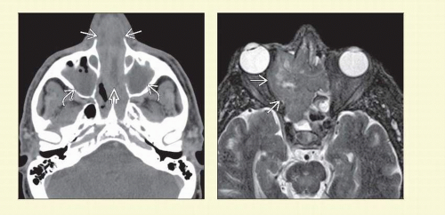 Figure 1 (Left) Axial CECT shows a large NHL  centered in the nasal cavity with slightly heterogeneous enhancement. There is gross destruction of the nasal septum centered in the nasal cavity with slightly heterogeneous enhancement. There is gross destruction of the nasal septum  . Obstructed secretions . Obstructed secretions  are noted in both maxillary sinuses. (Right) Axial STIR MR shows a large lymphoma involving the nasal cavity and ethmoid sinuses. Hypointense long TR signal is characteristic of this tumor with high nuclear to cytoplasmic ratio. Note the involvement of the right orbit are noted in both maxillary sinuses. (Right) Axial STIR MR shows a large lymphoma involving the nasal cavity and ethmoid sinuses. Hypointense long TR signal is characteristic of this tumor with high nuclear to cytoplasmic ratio. Note the involvement of the right orbit  with resulting proptosis. with resulting proptosis. |
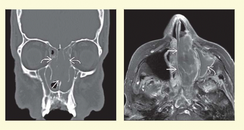 Figure 2 (Left) Coronal bone CT shows the classic location of sinonasal non-Hodgkin lymphoma. The mass is centered around the nasal septum, and there is destruction of the septum  as well as multiple ethmoid septations as well as multiple ethmoid septations  . (Right) Axial T1WI C+ FS MR demonstrates a large lymphoma filling the left nasal cavity. The septum is displaced . (Right) Axial T1WI C+ FS MR demonstrates a large lymphoma filling the left nasal cavity. The septum is displaced  but not eroded. Homogeneous mild enhancement is present. Trapped secretions but not eroded. Homogeneous mild enhancement is present. Trapped secretions  are present in the left antrum. are present in the left antrum. |
TERMINOLOGY
Abbreviations
Sinonasal non-Hodgkin lymphoma (NHL-SN)
Diffuse large B-cell lymphoma (DLBCL)
Natural killer/T-cell lymphoma (NKTL)
Definitions
NHL-SN: Extranodal lymphoproliferative malignancy most often of B-cell, T-cell, or NK/T-cell origin
NKTL: Subtype of peripheral T-cell lymphoma
NKTL previously called lethal midline granuloma, polymorphic reticulosis, angiocentric T-cell malignant lymphoma
IMAGING
General Features
Best diagnostic clue
Homogeneous soft tissue mass with predilection for nasal cavity ± bone destruction
Very nonspecific imaging features
NHL-SN can mimic variety of neoplasms & aggressive inflammatory disorders
Location
Nasal cavity > maxillary > ethmoid > frontal sinuses
NKTL may have simultaneous involvement of nasopharynx & oropharynx in addition to sinonasal cavities
Size
Usually between 2-5 cm
Morphology
Variable: Diffusely infiltrative & ill-defined, nodular, or bulky mass
CT Findings
NECT
Bulky, lobular, soft tissue mass in nasal cavity ± sinuses
May be hyperdense compared to soft tissue due to high nuclear-to-cytoplasmic (N:C) ratioRelated posts:

Stay updated, free articles. Join our Telegram channel

Full access? Get Clinical Tree


