A. Forces
–The mass of a body is a measure of its resistance to acceleration.
–Mass is measured in kilograms (kg).
–Velocity is the speed of a body moving in a given direction.
–Velocity is measured in meters per second (m/s).
–Acceleration is the rate of change of velocity.
–Acceleration is measured in meters per second squared (m/s2).
–A force causes a body to deviate from a state of rest or constant velocity (push or pull).
–Force = mass × acceleration, measured in newtons (N).
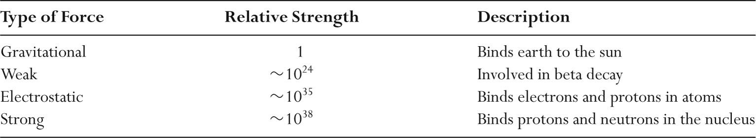
–The four physical forces in the universe are gravitational, electrostatic, strong, and weak.
–Relative strengths of these four forces are listed in Table 1.1.
–Gravity pulls objects to the Earth, and is important in cosmology.
–At the atomic level, effects of gravity are extremely small and are ignored.
–The electrostatic force causes protons and electrons to attract each other.
–Electrostatic forces hold atoms together.
–Strong forces hold the nucleus together.
–Weak forces are involved in beta decay.
B. Energy
–Energy is the ability to do work.
–Energy is measured in joules (J).
–Energy takes on various forms including electrical, nuclear, chemical, and thermal.
–One common form of energy is kinetic energy (KE) caused by motion.
–A bullet with mass m and velocity v has a kinetic energy of ½ mv2 .
–Another form of energy is potential energy (PE), which is the energy of position.
–A raised ball has potential energy.
–Energy cannot be created or destroyed.
–When a ball is released at a height, potential energy is converted into kinetic energy as the ball’s velocity increases.
–Einstein showed that mass and energy are interchangeable.
–E = mc2 where E is energy, m is mass, and c is the velocity of light.
–Rest mass energy is the energy equivalence of a particle.
–In diagnostic radiology, the electron volt (eV) is a convenient unit of energy.
–1 eV = 1.6 × 10⁻¹⁹ J
–One electron volt (1 eV) is the kinetic energy gained by an electron when it is accelerated across an electric potential of 1 volt (V) as depicted in Figure 1.1.
–An electron gains 1,000 eV (1 keV) when accelerated across an electric potential of 1,000 V.
–An electron gains 1 MeV (1,000 keV) when accelerated across an electric potential of 1,000,000 V.
–1 MeV = 103 keV = 106 eV
C. Electricity
–Electrons are negatively charged and protons are positively charged.
–Electric charge of an electron (or proton) is 1.6 × 10⁻¹⁹ coulomb (C).
TABLE 1.1 Relative Strength of Physical Forces
–Applying a voltage in an electrical circuit causes electrons to move.
–The positive region of an electrical circuit is called the anode.
–The negative region is called the cathode.
–Electrons are repelled from the cathode and attracted to the anode.
–Any voltage source in a complete circuit results in a flow of electrons in the circuit.
–Electric current, measured in amperes (A), is the flow of electrons through a circuit.
–An ampere is the amount of charge that flows divided by time.
–1 ampere = 1 coulomb per second
–Power supplies in any domestic home have a minimum of two wires and are single phase.
–Single-phase power supplies have one wire that has an oscillating voltage, with the other carrying no voltage.
–If there is a third wire, this is an “earth connection’’ for safety.
–In the United States, the electric power supply from utility companies is normally 110 volts (V).
–U.S. electricity is an alternating current (AC) that oscillates at a frequency of 60 cycles per second (60 Hz).
–In Britain, AC voltage is 220 V and oscillates at a frequency of 50 cycles per second (50 Hz).
–Three-phase power supplies have three lines of voltage, each 120 degrees out of phase with the others.
–Three-phase power supplies provide much more power than single phase.
D. Power
–Power is the rate of performing work.
–Power is the energy used divided by time, measured in watts (W).
–1 watt = 1 joule per second
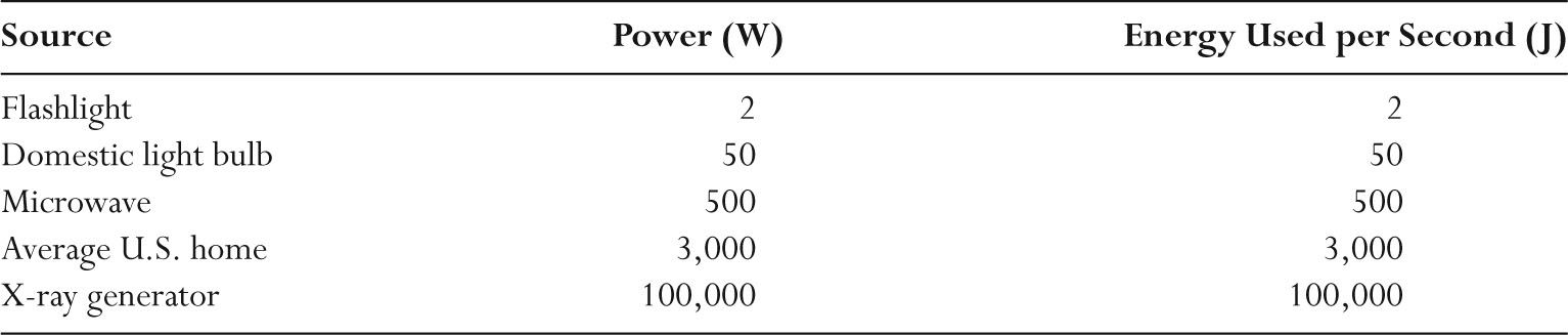
–Table 1.2 lists the power and energies of a range of sources.
–1 horsepower (HP) corresponds to 750 W.
–In electric circuits, the power (P) dissipated is the product of electric current (I) and voltage (V).
–Power (watt) = current (ampere) × voltage (volt)
–If the voltage is 100,000 V (100 kV) and the current is 1 A (1,000 mA), the power dissipated is 100,000 W (100 kW).
–A typical household in North America uses a few kW of electrical power.
–X-ray generators use up to 100 kW of electrical power, or the power required for ~30 U.S. households.
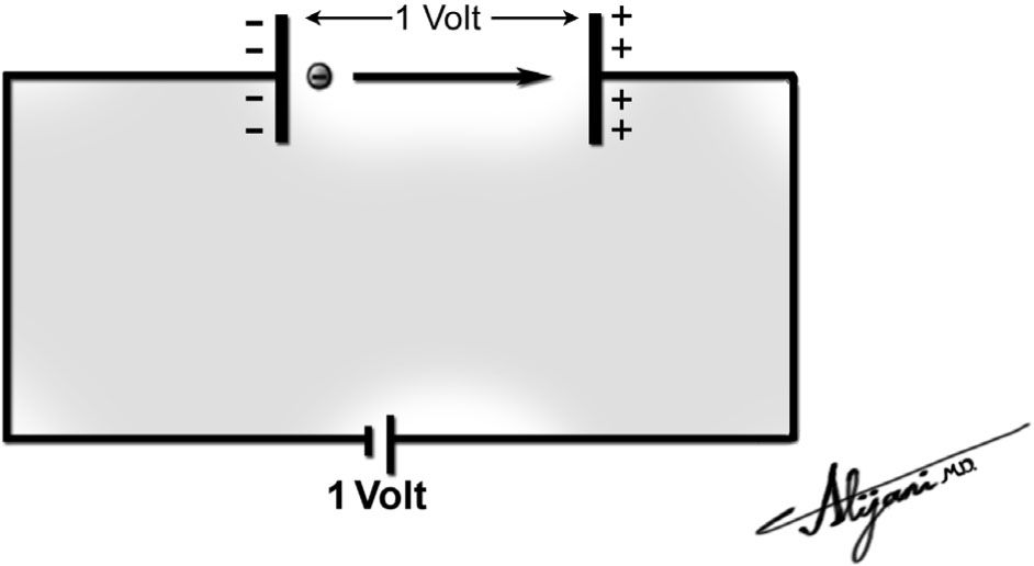
FIGURE 1.1 At the negatively charged plate, the electron has potential energy of 1 eV, which is converted into a kinetic energy of 1 eV as the electron is accelerated from the cathode to the anode.
TABLE 1.2 Common Power Sources
–The total energy generated is the product of power and time.
–Energy (joule) = power (watt) × time (second)
–X-ray generators are only switched on for short periods of time.
–A typical exposure time for a chest x-ray examination is 10 ms.
–Energy utilization in making x-rays is therefore low because of the very short exposure times that are used.
A. Waves
–A wave is an entity that varies in space and time.
–A common example of a wave is the variation of the water level in the ocean.
–Waves are characterized by a wavelength, frequency, and velocity.
–Wavelength (λ) is the distance between successive crests of waves.
–Wavelengths are measured in meters (m).
–Frequency (f) is the number of wave oscillations per unit of time.
–Frequencies are measured in cycles per second, where one cycle per second is equal to one hertz (Hz).
–The wave period is the time required for one wavelength to pass.
–Wave period is 1/f.
–The wave velocity (v) is the product of the wavelength and frequency, and measured in meters per second (m/s).
–Velocity (m/s) = frequency (Hz) × wavelength (m)
–Electromagnetic radiation is a wave that is associated with oscillating electric and magnetic fields.
–Visible light is a form of electromagnetic radiation.
–The sun emits (loses) the energy that it generates in nuclear processes by radiating visible light.
B. X-rays
–X-rays are a form of electromagnetic radiation.
–Electromagnetic radiation represents a transverse wave, in which the electric and magnetic fields oscillate perpendicular to the direction of the wave motion.
–Electromagnetic radiation travels in a straight line at the speed of light (c).
–The value of c is 3 × 10⁸ m/s in a vacuum.
–The product of the wavelength (λ) and frequency (f) of electromagnetic radiation is equal to the speed of light (c = fλ).
–Low-frequency electromagnetic radiation has a long wavelength.
–High-frequency electromagnetic radiation has a short wavelength.
–Figure 1.2 shows the electromagnetic spectrum that ranges from radio waves to gamma rays.
C. Photons
–Electromagnetic radiation is quantized, meaning that it exists in discrete quantities called photons.
–Photons may behave as waves or particles but have no mass.
–Photon energy (E) is directly proportional to frequency.
–Photon energy is inversely proportional to wavelength.
–Photon energy is E = h f = h (c/λ), where h is Plank’s constant.
–A 10-keV photon has a wavelength of 0.1 nm, comparable to the size of a small atom.
–A 100-keV photon has a wavelength of 0.01 nm.
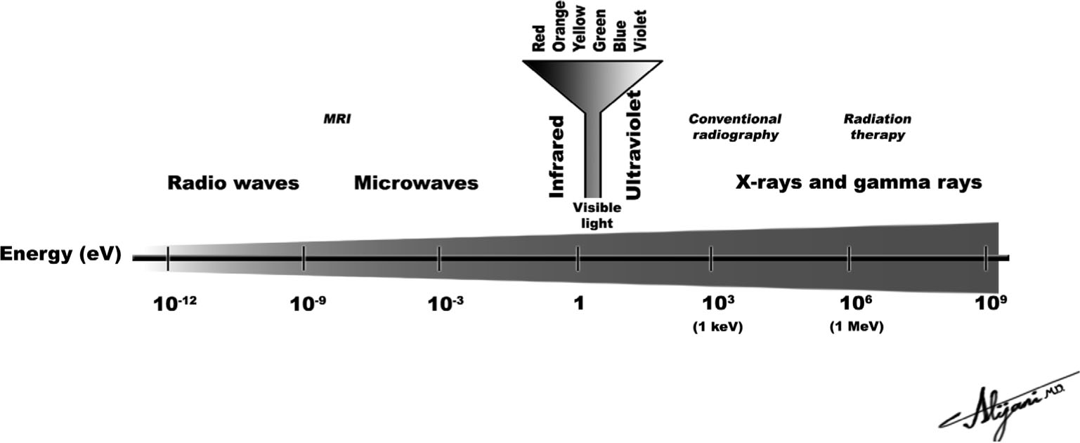
FIGURE 1.2 Electromagnetic spectrum ranging from radio waves to gamma rays showing that the photon energy is directly proportional to frequency.
–Radio waves have low frequencies (low photon energies) and gamma waves have high frequencies (high photon energies) as depicted in Figure 1.2.
–High energy photons are called x-rays if produced by electron interactions but gamma rays if produced in a nuclear process.
–There are no physical differences between x-rays and gamma rays of the same energy.
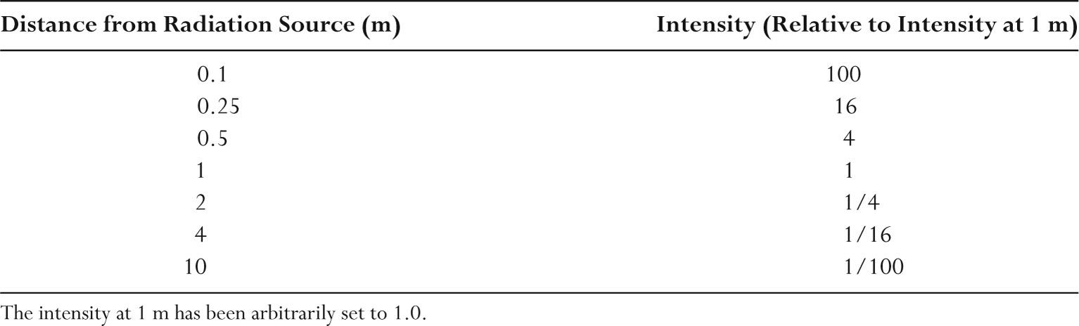
D. Inverse square law
–The intensity of an x-ray beam is proportional to the number of photons crossing a given area (e.g., square millimeter).
–X-ray beam intensity decreases with distance from the x-ray tube because of the divergence of the x-ray beam.
–The decrease in intensity is proportional to the square of the distance from the source. This nonlinear falloff in intensity with distance is called the inverse square law.
–Doubling the distance from the x-ray source decreases the x-ray beam intensity by a factor of 4.
–Halving the distance increases the x-ray beam intensity by a factor of 4.
–Table 1.3 shows how increasing and decreasing the distance from a source of radiation changes the radiation intensity.
–In general, if the distance from the x-ray source is changed from x1 to x2, then the x-ray beam intensity changes by (x1/x2)2.
A. Generator role
–X-ray generators provide electrical power to the x-ray tube.
–A small fraction of this power (~1%) is converted into x-rays.
–Virtually all current x-ray generators in Radiology departments use three-phase power supplies.
TABLE 1.3 Relative Radiation Intensity as a Function of Distance from the Radiation Source (Inverse Square Law)
–A generator uses a transformer to increase the voltage that is applied across the x-ray tube.
–The generator also rectifies the waveform from AC to direct current (DC).
–Generators permit x-ray operators to control three key parameters of x-ray operation.
–The tube voltage (kV) that is applied across the x-ray tube.
–The current (mA) that flows through the x-ray tube.
–The total exposure time (seconds) for which the tube current flows.
–The power dissipated equals the product of tube voltage (V) in volt and current (I) in amps, or VI, and is measured in watts (kW).
–Typical transformer ratings in x-ray departments are 100 kV and 1,000 mA, which correspond to a power of 100 kW.
B. Generator types
–Generators consist of an input power supply, transformer, and rectification circuit.
–Single-phase generators use a single-phase power supply.
–Single-phase generators use a bridge rectifier circuit that directs the alternating flow of high-voltage electrons so that flow is always from cathode to anode.
–Single-phase generators have been replaced by three-phase generators for use in diagnostic radiology.
–Single-phase generators are common for dental radiography where teeth are relatively thin and longer exposure times are tolerable (no moving parts).
–Three-phase generators use a three-phase power supply.
–High-frequency inverter generators transform an AC input into low-voltage DC, then into high-frequency AC, and finally into high-frequency AC waveforms that are rectified to yield a nearly constant voltage waveform.
–High-frequency generators are smaller and more efficient than three-phase generators.
–Constant potential generators provide a nearly constant voltage across the x-ray tube.
–Constant potential generators are expensive, require more space, and are used in interventional radiology.
C. Transformers
–A transformer changes the size of the input voltage and is capable of producing high and low voltages.
–Step-up transformers increase the voltage.
–A step-down transformer decreases the voltage.
–If two wire coils are wrapped around a common iron core, current in the primary coil produces a current in the secondary coil by electromagnetic induction.
–The voltages in the two circuits (Vp and Vs) are proportional to the number of turns in the two coils (Np and Ns)
–Np/Ns = Vp/Vs, where p refers to the primary and s to the secondary coils
–For an ideal transformer, the power in the primary and secondary circuits will be equal.
–VpIp = VsIs
–The step-up transformers used in x-ray generators have a secondary coil with many more turns (500:1) to produce a high voltage, which is applied across the tube.
–Generators also have a step-down transformer with fewer turns in the secondary coil.
–The step-down transformer produces a low voltage (10 V), which is applied across the x-ray tube filament circuit.
–An autotransformer permits adjustment of the output voltage using movable contacts to change the number of windings in the circuit.
D. Rectification
–The electric current from an AC power supply flows alternately in both directions, resulting in a voltage waveform shaped like a sine wave.
–Rectification changes the AC voltage into a DC voltage across the x-ray tube.
–Rectification is achieved using diodes, which permit current to flow in only one direction.
–Rectification for single-phase power supply normally uses four diodes and is called full-wave rectification.
–In full-wave rectification, there are two pulses per cycle of 1/60 second.
–AC electricity oscillation is 60 cycles per second.
–Each pulse ranges from zero volts to a peak (maximum) voltage.
–The maximum voltage is known as the kVp (p stands for peak).
–Rectification circuits in three-phase power supplies use a large number of diodes.
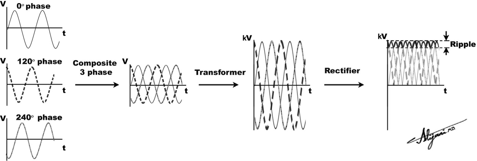
FIGURE 1.3 A three-phase generator (left) transforms and rectifies the input voltage to produce a high output voltage.
–Three-phase rectification circuits are arranged in combinations of delta and wye circuits.
–Many three-phase power supplies generate waveforms that have either 6 or 12 pulses per cycle of 1/60 second.
E. Voltage waveform
–Voltage waveform is a plot of voltage over time.
–A constant high voltage is desired across the x-ray tube for x-ray production.
–In practice, there is some variation in the voltage called ripple.
–The peak voltage or kilovolt peak (kVp) is the maximum voltage that crosses the x-ray tube during a complete waveform cycle.
–The voltage waveform ripple is the maximum voltage minus the minimum voltage per cycle expressed as a percentage of the maximum voltage.
–Single-phase systems have 100% ripple.
–Three-phase 6-pulse systems have ~13% ripple.
–Three-phase 12-pulse systems have ~4% ripple.
–High-frequency generators have ripple comparable to 12-pulse systems.
–Figure 1.3 shows how the waveform is created for a three-phase generator, and the corresponding ripple.
–The average (or effective) voltage will be slightly lower than the peak voltage.
–Most ripples in diagnostic radiology are relatively small (<10%).
–Nowadays in diagnostic radiology, kV and kVp are numerically similar. (For this reason, kV rather than technically correct kVp is used throughout this book.)
–When the ripple is high (e.g., 100%), it is important to differentiate the peak voltage from the average value, as the latter will be much lower than the applied kVp.
Related posts:


Stay updated, free articles. Join our Telegram channel

Full access? Get Clinical Tree


