Case 1
 Clinical Presentation
A 62-year-old woman with gross hematuria.
 Imaging Findings
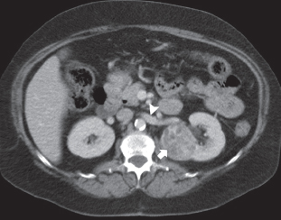
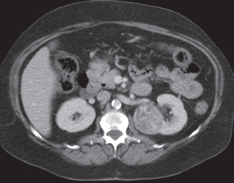
The contrast-enhanced computed tomography (CT) sections through midabdomen show a heterogeneously enhancing mass (arrow) occupying the interpolar region of the left kidney. The mass is centered in the renal parenchyma and causes contour deformity of the kidney. It is not surrounded by a dilated collecting system. No involvement of neighboring structures is seen. No lymphadenopathy is seen in these sections. Left renal (arrowhead) vein is well seen and shows no enlargement or obvious filling defect.
 Differential Diagnosis
• Renal cell carcinoma (RCC): Heterogeneous enhancement of the mass and origin from the renal parenchyma are findings characteristic of RCC.
• Oncocytoma: This also presents as an enhancing mass arising from the renal parenchyma. A spoke wheel pattern of enhancement is considered characteristic. However, because it cannot be reliably distinguished from RCC and is much less common than that entity, oncocytoma should not be diagnosed prospectively
• Lipid-poor angiomyolipoma:
Stay updated, free articles. Join our Telegram channel

Full access? Get Clinical Tree


