Kaye D. Westmark, Laura Ocasio, and Roy F. Riascos
1.1 Case Presentation
1.1.1 History and Physical Examination
A 45-year-old man presented with a history of migraine headaches which had recently worsened in severity.
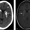
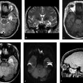
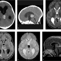
His neurological examination was normal. Specifically, cranial nerves II to XII were intact. An axial T2-weighted image from his brain MRI is shown in ▶ Fig. 1.1.
Fig. 1.1Fig. 1.2(a) The axial FSE T2-weighted image reveals a vascular-appearing flow void (arrow), extending from the right cavernous sinus into the prepontine cistern. (b) This abnormal vessel is confirmed on the COW MRA, where the proximal basilar artery appears small and the anomalous vessel, arising from the cavernous portion of the right internal carotid artery (arrow), can be seen contributing to the distal basilar artery, which it joins immediately proximal to the origin of the superior cerebellar arteries. The P-com arteries were hypoplastic bilaterally. These findings are consistent with a persistent primitive trigeminal artery. COW, circle of Willis; FSE, fast spin echo; MRA, magnetic resonance angiography; P-com, posterior communicating.
Only gold members can continue reading. Log In or Register to continue