9 Hyperostosis Frontalis Interna
9.1 Case Presentation
9.1.1 History and Physical Examination
A 45-year-old woman presents with headache and nausea.
Physical examination was normal with no neurologic deficit.
9.2 Differential Diagnosis
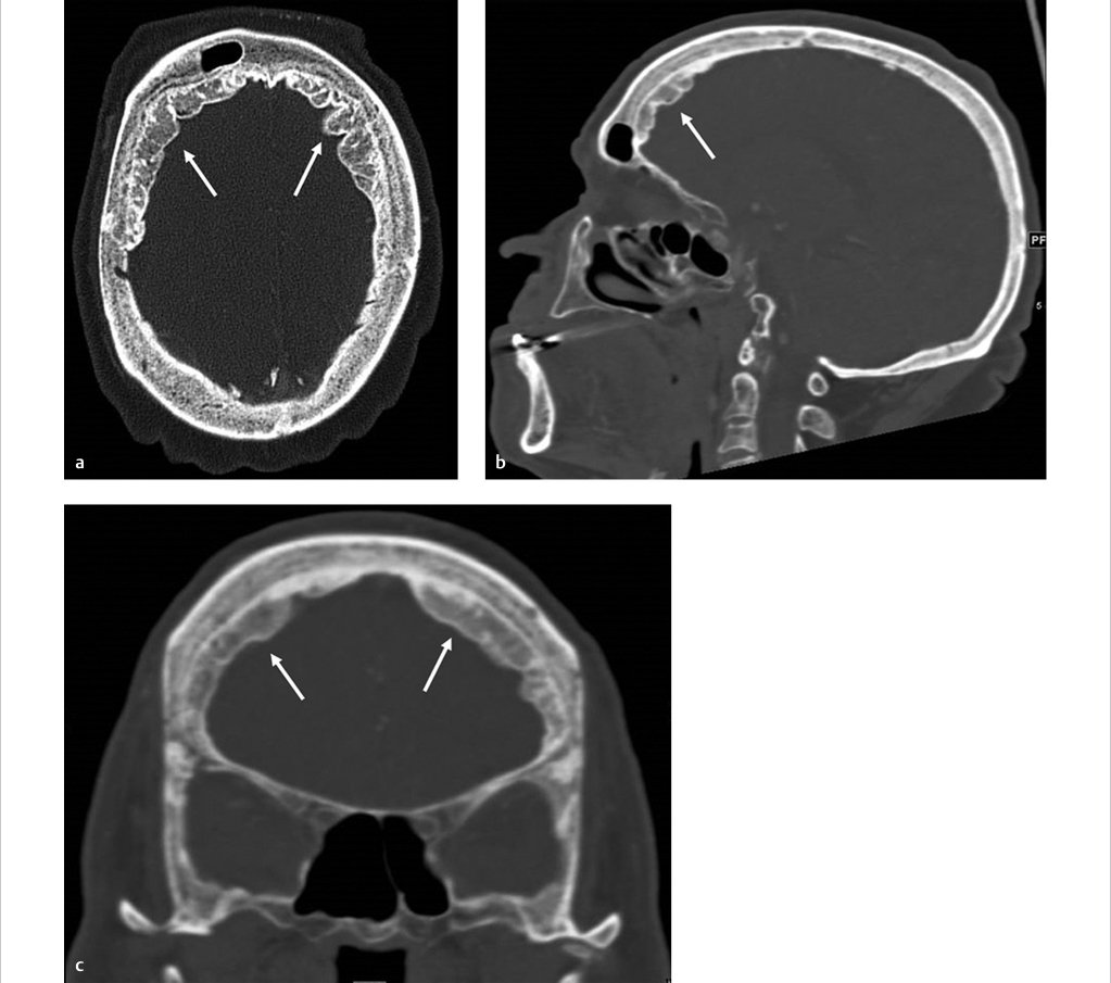
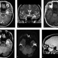
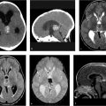
Hyperostosis frontalis interna:
Symmetrical thickening involving the inner table of the frontal bones with no enhancement. It is frequently seen in middle-aged women.
Acromegaly:
Diffuse calvarial thickening associated with enlargement of the sinuses, enlargement of the sella turcica, frontal bossing, and prominence of the mandible.
Other skeletal features are also present involving the spine and joints.
Fibrous dysplasia:
A non-neoplastic condition involving the bones and commonly occurring in children and young adults.
Normal bone is replaced with fibrous stroma and immature woven bone. The skull is involved in the polyostotic form.
It is characterized by focal or diffuse ground glass thickening of the skull.
The ethmoid bone is the most common involved area of the skull base.
The inner table is more frequently involved.
Paget’s disease of the bone:
A chronic bone disorder characterized by excessive bone remodeling with replacement of normal bone architecture with a coarsened trabecular pattern.
Commonly involved sites include the skull, spine, pelvis, and long bones.
It is more common in individuals older than 40 years with a slight male predilection.
Involvement of the skull is characterized by diffuse thickening of the inner and outer tables as opposed to fibrous dysplasia, which predominantly affects the inner table.
Other findings in the skull are widening of the diploic space, mixed lytic and sclerotic lesions (cotton wool appearance), or large well-defined lytic lesions (osteoporosis circumscripta).
Enhancement of the lesions is seen in active disease.
Meningioma:
An extra-axial tumor arising from the arachnoid cap cells of the meninges.
Classically, it presents as a homogeneously enhancing dural-based lesion with a characteristic dural tail sign.
It is often associated with hyperostosis of the overlying skull that has been shown to be due to either hypervascularity or direct infiltration of tumor cells.
Sclerotic metastasis:
It can arise from a variety of primary malignancies including prostate carcinoma, breast carcinoma, transitional cell carcinoma of urothelial origin, medulloblastoma, neuroblastoma, and lymphoma, among others.
It is characterized by single or multiple focal areas of increased density as compared to the normal skull.
It tends to enhance.
Related posts:

Stay updated, free articles. Join our Telegram channel

Full access? Get Clinical Tree


