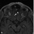
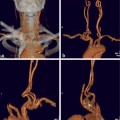
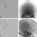
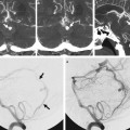
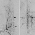
Fig. 1.1 Contrast-enhanced MRA of the arch and cervical vessels (a) and LCCA angiogram in anteroposterior (AP) view at the arch and neck (b) demonstrate the common origin of the brachiocephalic trunk and the LCCA from the aortic arch — the so-called “bovine arch.” There are multifocal stenoses of the LCCA. Reflux of contrast from the origin of the LCCA into the common trunk and into the RCCA is observed. Note the signal gap in the MRA resulting from local field inhomogeneity related to the stent (within the RCCA).
1.1.3 Diagnosis
Multifocal postradiation stenosis in a patient with a common supra-aortic trunk that gives rise to the brachiocephalic trunk and the LCCA.
1.2 Embryology and Anatomy
Development of the aortic arch and great vessels results from formation and selective involution of paired vascular arches connecting the embryonic aortic sac (ventral aorta) with the paired dorsal aortae.
Between the second and seventh week of gestation, branchial apparatus development occurs, with formation of six paired branchial arches in the wall of the foregut, numbered from cephalad to caudad. The embryonic aortic vascular arches arise in a cranial to caudal sequence, forming primitive arterial arcades around the pharyngeal arches. Regression or disappearance of some vascular arch segments, with persistence and remodeling of others, results in the formation of the thoracic aorta and its major branches. The first and second pair of vascular arches appear by days 24 and 26, respectively; the third and fourth by day 28; and the sixth by day 29. The fifth arch never fully develops or appears briefly and then involutes. By the time of appearance of the sixth arch, the first and second arches have largely regressed.
The ascending thoracic aorta is formed from the truncus arteriosus proximally and the aortic sac/ventral aorta distally. The aortic sac forms right and left horns. The right horn subsequently gives rise to the brachiocephalic artery, whereas the left forms the proximal portion of the aortic arch between the brachiocephalic arteries and LCCAs. The ventral aorta and the third vascular arches form paired primitive carotid arteries from which intracranial blood flow during early development is primarily derived. The CCAs are derived from the proximal portions of these primitive vessels.
The segment of the dorsal aorta between the third and fourth arches obliterates, and the fourth and sixth vascular arches undergo asymmetric remodeling. On the left side, the fourth arch and dorsal aorta form the definitive aortic arch (between the LCCA and left subclavian arteries) and the most proximal portion of the descending thoracic aorta. On the right side, the fourth arch and part of the dorsal aorta constitute the proximal portions of the right subclavian artery, with gradual loss of connection to the midline descending aorta. The ventral portions of the sixth vascular arches give rise to the pulmonary arteries. The dorsal contribution of the sixth arch regresses on the right while persisting on the left side as the ductus arteriosus.
Stay updated, free articles. Join our Telegram channel

Full access? Get Clinical Tree


