Even in patients who are on mechanical ventilation, obstructive atelectasis can occur very rapidly. One theory is that highly oxygenated air, as opposed to normal ambient air, is quickly absorbed into the alveolar capillaries from the alveoli, so that collapse occurs within minutes.
Passive atelectasis is very often encountered in the ICU patient due to pleural effusions. Pneumothorax is also a cause of passive atelectasis. The lung tends to collapse on itself especially in the most dependent regions, where the effects of gravity help the effusion compress the lung. Thus partial lobar consolidations are more often seen in the lower lobe, particularly on the left, and are seen only occasionally in the upper lobes. The radiographic hallmark of this collapse is disappearance of the diaphragm, and it is important to differentiate atelectasis from effusion. Look for the presence of an air bronchogram, which is typical of this type of atelectasis. A CT will also demonstrate a homogeneous consolidation with a central air bronchogram.
Compressive atelectasis may be seen secondary to a preexisting mass, either a tumor or an abscess. These causes are somewhat unusual and the most common cause of compressive atelectasis is pleural fluid.
Many areas in the lung are can become atelectatic, but the left lower lobe is by far the most common site. Atelectasis of the right lower lobe occurs about a third less often than in the left lower lobe, and collapse of the right upper lobe occurs approximately 50% less than in the right lower lobe.
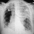
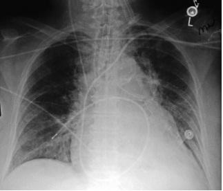
Collapse of the left lower lobe may be due to phrenic nerve injury, but most often occurs due to an enlarged left atrium, which compresses the left lower lobe bronchus (Fig. 10.2).139
Figure 10.2 A portable film reveals an enlarged heart with left atrial enlargement. Note the atelectasis and collapse of the left lower lobe. The Swan-Ganz catheter is too far out into the right lower lobe bronchus.
Cicatricial atelectasis is volume loss secondary to pulmonary fibrosis and is most commonly seen in patients with underlying pulmonary disease. Often, this is seen in patients who develop significant pulmonary fibrosis as a late complication of ARDS.
The radiographic appearance of atelectasis depends on the extent and the location of the volume loss. Peripheral subsegmental atelectasis presents as linear densities predominately in the lung bases. This produces the so-called linear or plate-like atelectasis (Fig. 10.3).
Stay updated, free articles. Join our Telegram channel

Full access? Get Clinical Tree


