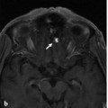
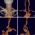
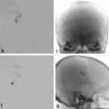
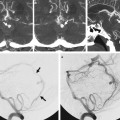
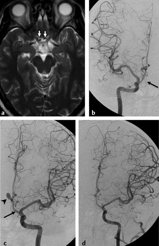
The Infraoptic Course of the Anterior Cerebral Artery A 34-year-old man presented with anxiety-related episodic hypertension. On physical examination, he was found to have a heart murmur and congenital absence of the ring and little finger of his left hand. He was subsequently diagnosed with having a bicuspid aortic valve and postductal coarctation of the aorta, which was treated successfully with aortic stenting. Screening cerebral magnetic resonance angiography showed an infraoptic course of bilateral anterior cerebral arteries (ACAs) and a 7.5-mm incidental anterior communicating artery (AcomA) aneurysm. See ▶ Fig. 10.1. Fig. 10.1 T2-weighted MRI (a) shows an infraoptic course of the A1 segments of the ACAs bilaterally, with an abnormal medial course of the A1 segments relative to the optic nerves (white arrows). Right (b) and left ICA (c) angiograms in anteroposterior views show the infraoptic course of the A1 segments of bilateral ACAs (arrows). There is an outpouching at the left A1–A2 junction, representing a left AcomA aneurysm (arrowhead). Left ICA angiogram postcoiling in anteroposterior view (d) reveals complete occlusion of the left AcomA aneurysm after coiling. Left AcomA aneurysm with infraoptic course of bilateral ACAs. During normal embryogenesis, the rostrolateral portion of the perioptic arterial plexus gives rise to the terminal segment of the internal carotid artery (ICA) and the primitive ophthalmic artery. The middle cerebral, anterior cerebral, posterior communicating, anterior choroidal, and temporary dorsal and ventral ophthalmic arteries arise at this site. The embryogenesis of an infraoptic ACA is still controversial. Some of the hypotheses proposed in the literature are that: the embryonic anastomosis between the primitive maxillary artery and the ACA may persist; an anomalous vessel may arise from the persistent enlarged embryonic anastomotic loop between primitive dorsal arteries and ventral ophthalmic arteries; the primitive prechiasmal anastomosis between the branches of the ICA, ophthalmic artery, and ACAs may persist. The error in embryogenesis is likely to occur in the early stages of cranial arterial development at the level of the perioptic arterial plexus. The characteristic features of an infraoptic course of the A1 are that the artery arises from the ICA at the level of the ophthalmic origin and is located underneath the optic nerve to supply distally the territorial distribution of a normal ACA via an anastomotic segment in the vicinity of the ACA. Approximately 75% of infraoptic ACAs are seen on the right side, 15% on the left side, and 10% bilaterally. The configurations of proximal ACAs in the presence of an infraoptic A1 have been classified into four types, as demonstrated in ▶ Fig. 10.2.
10.1 Case Description
10.1.1 Clinical Presentation
10.1.2 Radiological Studies
10.1.3 Diagnosis
10.2 Embryology and Anatomy
Stay updated, free articles. Join our Telegram channel

Full access? Get Clinical Tree


