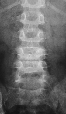
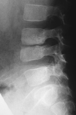
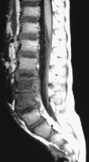
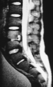
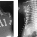
CASE 100 A 5-year-old boy presents with back pain and fever. Figure 100A Figure 100B Frontal and lateral radiographs of the lumbar spine demonstrate decreased intervertebral disk space height at L3-L4. The adjacent vertebral body end-plate margins are indistinct and irregular with underlying areas of sclerosis and erosions (Figs. 100A to 100B). Corresponding sagittal MRI show inhomogeneous decreased signal in the L3 and L4 vertebral bodies on T1-weighted imaging (Fig. 100C) with increased signal on T2-weighted imaging (Fig. 100D). There is narrowing of the L3-L4 disc space and retrograde protrusion of a portion of disc or fluid from inflammation. Figure 100D Diskitis with vertebral osteomyelitis. Diskitis and vertebral osteomyelitis are disease processes with distinct epidemiologic, clinical, and radiographic features. Diskitis is an inflammatory process of the disk space, more common in children than adults, especially in girls (2x). The disease affects the lumbar spine predominantly. The mean age at presentation is 2.8 years with bimodal age peaks at 6 months to 4 years and 10 to 14 years. Vertebral osteomyelitis most commonly also affects the lumbosacral region (64%) but can be seen in the thoracic spine (29%) and occasionally in the cervical location. The mean age at presentation for vertebral osteomyelitis is 7.5 years with the duration of symptoms longer on average than diskitis (33 days vs 22 days). Whether diskitis and vertebral osteomyelitis are distinct processes or reflect a spectrum of a single disease is controversial. Early in the course of the disease, differentiation clinically can be difficult. The imaging modality of choice in a patient with possible vertebral osteomyelitis is MRI, which has high sensitivity and specificity.
Clinical Presentation
Radiologic Findings
Diagnosis
Differential Diagnosis
Discussion
Background
Etiology
Related posts:


Stay updated, free articles. Join our Telegram channel

Full access? Get Clinical Tree


