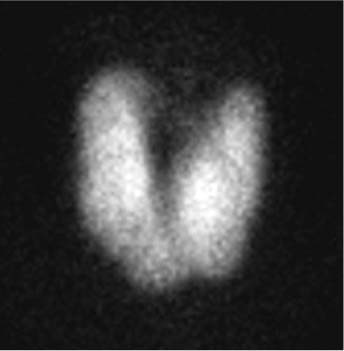
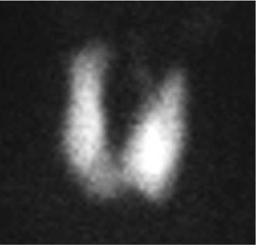
CASE 100 A 26-year-old woman presents with clinical and biochemical evidence of hyperthyroidism. Fig. 100.1 • 99mTc-pertechnetate, 10 mCi (370 MBq) intravenously. • Planar imaging of the neck with a pinhole collimator 20 minutes later (Figs. 100.1 and 100.2). Anterior view from the 99mTc-pertechnetate scan, with pinhole collimator 4 cm from the neck (Fig. 100.1), demonstrates intense, increased uptake throughout the substantially enlarged thyroid. The intense uptake in the thyroid results in little visualization of the background tissues. A pyramidal lobe is appreciated extending superiorly from the upper aspect of the left lobe. A 6-hour radioiodine uptake assessment earlier the same day revealed a markedly elevated uptake of 80% (normal, 5–20%). The up-take and scan findings are consistent with Graves disease. An oral dose of 13 mCi of 131I was administered. The patient was still hyperthyroid 10 months later and was referred back to nuclear medicine. The repeated scan (Fig. 100.2) again demonstrated intense up-take throughout an enlarged thyroid, although it was smaller than on the initial study. There was again visualization of a pyramidal lobe and little visualization of the background tissues. Repeated 6-hour radioiodine uptake was 47%, significantly less than on the original study, but still elevated. She was retreated with 9 mCi of 131I, and her hyperthyroidism has not recurred. Fig. 100.2 The treatment of hyperthyroidism with 131
Clinical Presentation
Technique
Image Interpretation
Therapy
Discussion
![]()
Stay updated, free articles. Join our Telegram channel

Full access? Get Clinical Tree


