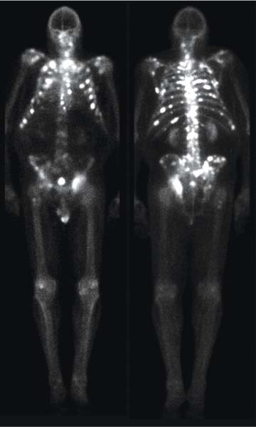
CASE 102 A 76-year-old man with prostate cancer and pain from bone metastases undergoes palliative external beam radiotherapy to his left hip with symptomatic relief. However, the symptoms recur and involve multiple sites despite optimal medical treatment. He is deemed not to be a candidate for further external beam radiotherapy because of the multifocal nature of his symptoms, and he is referred to nuclear medicine for systemic radioisotope therapy. Fig. 102.1 • 99mTc-MDP, 25 mCi (925 MBq) intravenously. Planar imaging with a low-energy, high-resolution collimator 3 hours later (Figs. 102.1 and 102.2A). • 89Sr (148 MBq) intravenously (therapy dose). The 99mTc-MDP bone scan (Fig. 102.1) reveals numerous sites of metastatic disease, predominantly within the axial skeleton. The patient was administered 89Sr, 4 mCi, by slow intravenous injection over 2 minutes. Following therapy, he became pain free for a period of about 8 months. His pain subsequently recurred. Given the favorable response to the initial 89Sr therapy, this treatment was repeated. In the later stages of malignancy, pain from bone metastases is a common symptom and often adversely affects the quality of life. When disseminated metastases result in multiple sites of bone pain, external beam radiotherapy is no longer practical. In this setting, radiotherapy with systemically administered radiopharmaceuticals has proved successful in alleviating bone pain. Originally performed with sodium phosphate 32P, the most commonly used agents today are: 89Sr, administered as strontium chlo-ride 89Sr; 153Sm, administered as 153Sm-EDTMP (ethylene diamine tetramethylene phosphonate); and less commonly, 186Re, administered as 186Re-etidronate (186
Clinical Presentation
Technique
Image Interpretation
Therapy
Discussion
![]()
Stay updated, free articles. Join our Telegram channel

Full access? Get Clinical Tree


