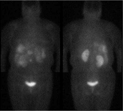
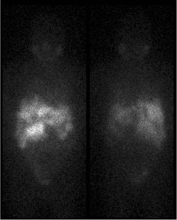
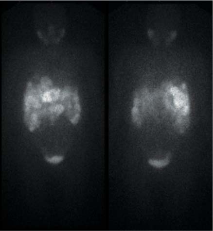
CASE 104 A 45-year-old woman presents with a carcinoid tumor and multiple liver metastases. Because of the multifocal nature of her disease and subacutely progressive symptoms despite optimal chemo-therapy, she is referred to Nuclear Medicine for consideration of systemic radioisotope therapy. Fig. 104.1 Fig. 104.2 • 111In-pentetreotide, 3 mCi (111 MBq) intravenously. Planar imaging with a medium-energy collimator 48 hours after injection (Fig. 104.1). • 131I-MIBG, 0.5 mCi (18 MBq) intravenously. Planar imaging with a high-energy collimator 48 hours after injection (Fig. 104.2). • 123I-MIBG, 6 mCi (222 MBq) intravenously. Planar imaging with a low-energy, high-resolution collimator 24 hours after injection (Fig. 104.3). The original 111In-pentetreotide scan (Fig. 104.1) reveals numerous areas of increased and decreased uptake throughout the liver, in keeping with active and necrotic carcinoid metastases, respectively. Because therapy with 131I-MIBG is being considered, it is necessary to demonstrate that all active tumor sites are MIBG-avid, and consequently a 131I-MIBG scan is performed (Fig. 104.2), confirming that this is the case. Fig. 104.3 The patient was administered a slow intravenous infusion of 150 mCi of 131I-MIBG without any immediate side effects. Her platelet count decreased from 215,000/mm3 before therapy to a nadir of 94,000/mm3 8 weeks later, then rebounded to 351,000/mm3 another 8 weeks later. She subsequently reported mild relief of her symptoms. A follow-up MIBG scan 3 months after therapy, this time labeled with 123I rather than 131I (because of a change in institutional procedure) demonstrated stability of her disease (Fig. 104.3). There was no significant change in size of the metastases on CT. She was then re-treated with 131I-MIBG as the next step in a multiple-dose regimen.
Clinical Presentation
Technique
Image Interpretation
Therapy
Discussion
Stay updated, free articles. Join our Telegram channel

Full access? Get Clinical Tree


