Case 11
 Clinical Presentation
A 48-year-old woman with an enlarged uterus.
 Imaging Findings
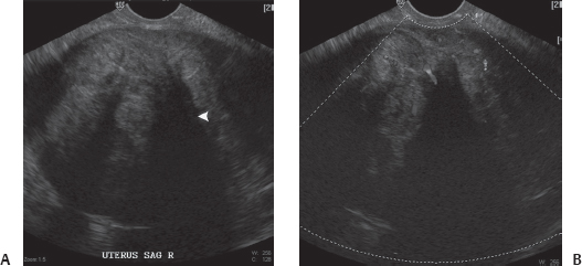
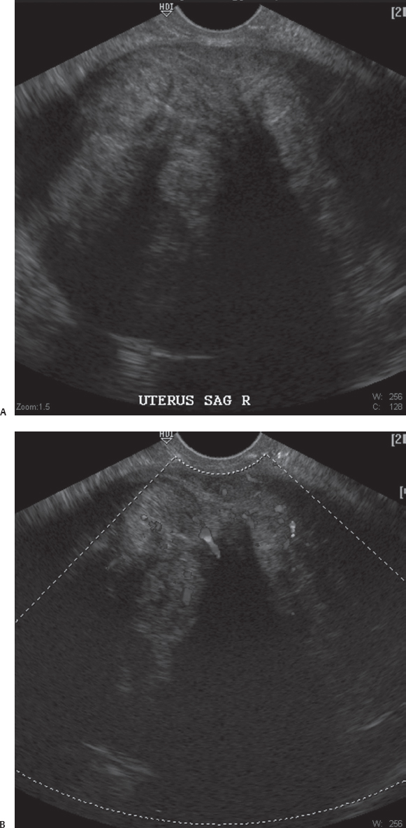
(A) Transvaginal sonographic image marked as “sagittal uterus” shows the uterus to be enlarged by a large hypoechoic mass with a whorled pattern, which shows some shadowing emanating from within its substance (arrowhead). (B) Transvaginal sonographic Doppler image at the same level as Figure A shows vascularity within the substance of the mass.
 Differential Diagnosis
• Uterine leiomyoma: A hypoechoic whorled pattern is characteristic of this diagnosis. The endometrial canal may be deformed by large lesions. Shadowing shows internal calcifications.
• Endometrioma: A large endometrioma may superficially resemble a large uterine leiomyoma. Uniform low-level internal echogenicity through the transmission area is a characteristic finding. No internal flow can be demonstrated.
• Solid ovarian neoplasm:
Stay updated, free articles. Join our Telegram channel

Full access? Get Clinical Tree


