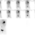
CASE 112 A 77-year-old woman on peritoneal dialysis has fever and abdominal pain. Fig. 112.1 Whole-body 4-hour planar image, anterior and posterior projections, 99mTc-WBCs. Fig. 112.2 Whole-body 24-hour planar image, anterior and posterior projections, 99mTc-WBCs. Fig. 112.3 abdominal 24-hour sPeCT, coronal projection, 99mTc-WBCs. • A 20 mCi dose of white blood cells labeled with 99mTc-WBCs is injected intravenously. • Whole-body planar imaging in anterior and posterior projections at 4 hours and 24 hours • Abdominal SPECT at 24 hours • Dual-detector gamma camera • Low-energy, high-resolution collimators • Energy peak of 140 keV At 4 hours, blood pool activity and early localization in the gallbladder fossa and left posterior pelvis are subtle findings (Fig. 112.1
Clinical Presentation
Technique
Image Interpretation
![]()
Stay updated, free articles. Join our Telegram channel

Full access? Get Clinical Tree


