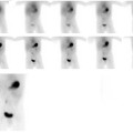
CASE 114 A 37-year-old woman is incidentally noted to have a 2.5-cm mass-like structure in the interpolar region of the right kidney on a gynecologic ultrasound examination. Follow-up MRI shows a 2.6 × 2.0-cm prominence in the right interpolar region that is equivocal for renal tumor versus hypertrophic columns of Bertin. Fig. 114.1 Fig. 114.2 Fig. 114.3 • Inject 1 to 5 mCi of 99mTc-DMSA. Use within 30 minutes of labeling, and avoid introducing air during the labeling reaction. • Use a low-energy, high-resolution collimator. • Image 2 to 3 hours after injection. • For infants, pinhole collimators may be used for better anatomic definition. For adults, SPECT is preferable, given the greater depth of the kidney. Dynamic images (Fig. 114.1) show prompt perfusion of both kidneys, which appear similar in size. On the 4-minute static image (Fig. 114.2), both kidneys show homogeneous uptake. Note that the blood pool activity persists longer than on 99mTc-DTPA or 99mTc-MAG-3 imaging because of slower cortical uptake. Figure 114.3 shows SPECT images (top) obtained at 3 hours after injection. Fusion with MR image (bottom) indicates that the mass-like feature adjacent to the right renal pelvis at the crossline intersection has 99mTc-DMSA uptake similar to that of the remainder of the renal cortex, confirming that it consists of functional renal tissue.
Clinical Presentation
Technique
Image Interpretation
Stay updated, free articles. Join our Telegram channel

Full access? Get Clinical Tree


