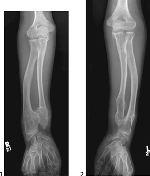
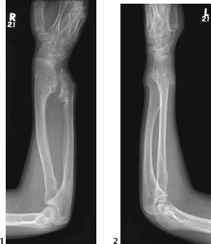
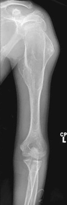
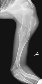
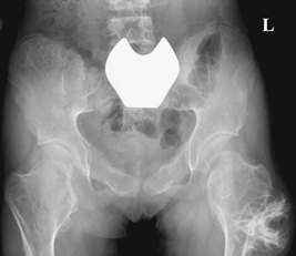
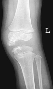
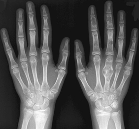
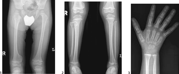
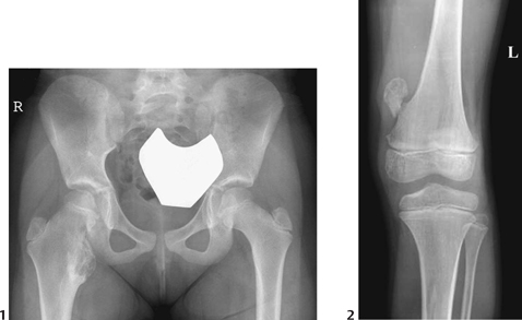
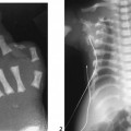
CASE 114 A young child presents with multiple bilateral painless, hard lumps adjacent to several joints. He also has mild limitation of wrist and elbow motion. Figure 114A Figure 114B Figure 114C Figure 114D Figure 114E The tubular bone and modeling deformities shown in Figs. 114A to 114E demonstrate metaphyseal broadening from multiple exostoses. Associated deformities of adjacent joints are present, including the distal radioulnar and radiocarpal joints. Note the variable appearance of the exostoses affecting the proximal humeri, femora, and ribs. Figure 114F Frontal radiograph of the knee of a patient with Trevor disease showing enlargement of the distal femoral and proximal tibial epiphyses medially with irregularity in ossification. Figure 114G Hand radiograph of a patient with Ollier disease showing multiple enchondromatosis with expansion of the medullary space especially involving the left third metacarpal. Figure 114H Radiographs of hip and femur (1), lower limbs (2), and right hand (3) of a patient with Langer-Giedion syndrome. Note the multiple exostoses of the right femur and fibula especially. Multiple hereditary exostoses (MHE) (osteochondromatosis, diaphyseal aclasis) Figure 114I Two examples of isolated exostosis involving the right lesser trochanter (1) and distal femur (2).
Clinical Presentation
Radiologic Findings
Diagnosis
Differential Diagnosis
Discussion
Background
Related posts:


Stay updated, free articles. Join our Telegram channel

Full access? Get Clinical Tree


