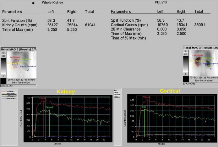
CASE 118 A 34-year-old woman with a history of upper respiratory infection/sore throat presents with abnormally elevated serum creatinine. Fig. 118.1 Fig. 118.2 • Inject 10 mCi of 99mTc-MAG-3 with a tight intravenous bolus. 99mTc-DTPA (10–20 mCi) can also be used. • Use a low-energy, medium-resolution collimator. • Image native kidneys in the posterior view. • Image for the first minute with 1- to 2-second frames. Then image for 2 to 30 minutes with 30- to 60-second frames. A 5-minute static image at the end may help to demonstrate ureteral uptake. Posterior dynamic images (Fig. 118.1) show a right kidney slightly smaller than the left, without focal abnormalities. The split function is 58% left and 42% right, likely reflecting this anatomic asymmetry. On the time–activity curves (Fig. 118.2), cortical uptake is relatively brisk, with maxima at 3.2 minutes on the left and 2.5 minutes on the right. Clearance is moderately delayed, however, and at the end of the study, residuals of 80% and 66% are seen on the left and right, respectively. • Glomerulonephritis, probably post-infectious by history • Acute tubular necrosis (mild) • Lupus nephritis • Drug-induced nephropathy • Diabetic nephropathy • Hypertensive nephropathy
Clinical Presentation
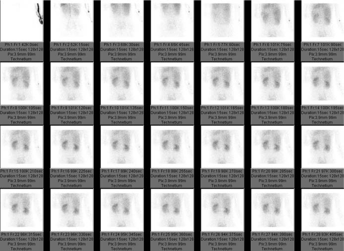
Technique
Image Interpretation
Differential Diagnosis
Diagnosis and clinical Follow-Up
Related posts:


Stay updated, free articles. Join our Telegram channel

Full access? Get Clinical Tree


