Diagnosis: Pseudomembranous colitis with hemorrhage into bowel lumen
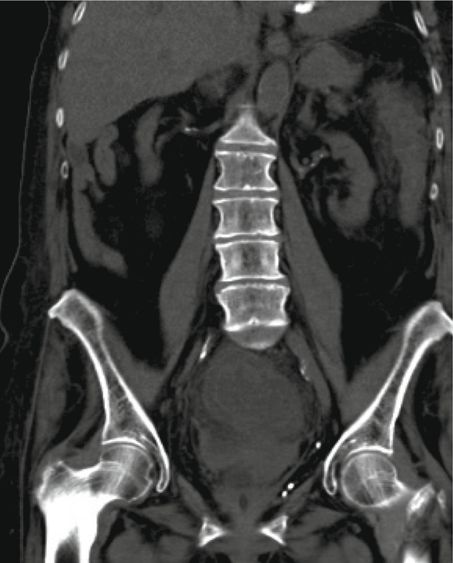
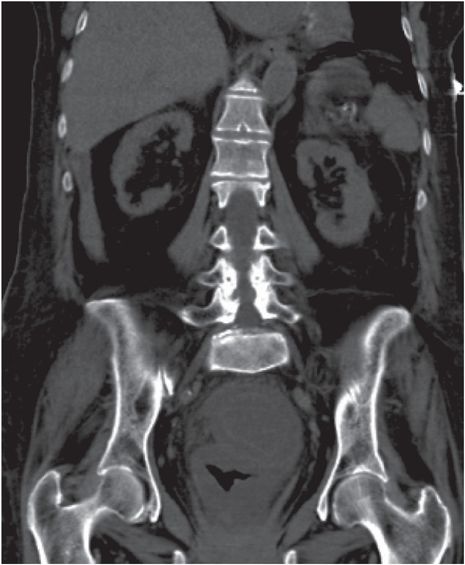
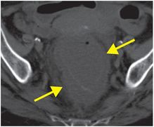
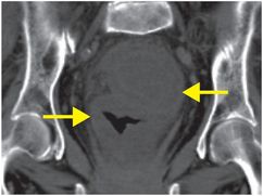
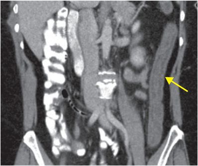
Axial (left image) and coronal (middle and right images) noncontrast CT images show a markedly thickened sigmoid colon (arrows), with distension of the lumen with high-attenuation fluid, representing intraluminal hemorrhage.
Discussion
Overview and imaging of colitis
Inflammation of the colon is a common cause of abdominal pain.
Several unrelated disease processes of infectious, ischemic, or inflammatory etiology can lead to a similar CT appearance of colonic wall thickening. Secondary imaging findings and clinical reasoning are often needed to narrow the differential diagnosis.
The hallmark CT imaging feature of colitis is bowel wall thickening, and most often, a final diagnosis requires a colonoscopic biopsy. However, there are several other findings that can help narrow the differential diagnosis.
For instance, in addition to evaluating the degree of colonic wall thickening, what is the pattern of involvement of the colon and the small bowel? Is there ascites? Is there an associated fluid collection? Is there atherosclerotic disease or thrombosis affecting the vasculature?
Incidental colonic wall thickening mimicking colitis can be found in greater than 10% of abdominopelvic CT scans.
Some authors recommend that any patient with colonic thickening seen on CT receive a follow-up colonoscopy to evaluate for diseases that would require ongoing management, such as colon carcinoma or inflammatory bowel disease.
Clinical synopsis
This patient, a 68-year-old male with hemorrhagic pseudomembranous colitis and hypotension, was admitted directly to the ICU for stabilization of blood pressure. He required emergent colectomy, and ultimately was discharged to a nursing home after a protracted hospital stay.
Self-assessment
|
|
|
|
Etiology of colitis
Infectious colitis
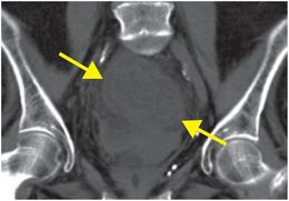
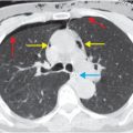
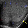
Several organisms cause colitis. Yersinia, salmonella, and tuberculosis tend to affect the right colon. Tuberculosis attacks the ileocecal valve with a desmoplastic reaction that may mimic Crohn’s disease. Imaging features are often nonspecific. In general, bacterial colitis features pericolonic stranding and ascites; however, stool studies or biopsy are necessary for a definitive diagnosis. Coronal CT demonstrates nonspecific thickening of the left colon (arrow) in this case of infectious colitis.
Related posts:
 8 67-year-old man with a history of altered mental status, left flank pain, and hypotension, on anticoagulant medications
 32 32-year-old male complaining of chest pain after upper endoscopy
 28 30-year-old male presented with a palpable left testicular mass
 29 19-year-old male presented with acute onset right scrotal pain
 53 42-year-old female presenting with fever and back pain
 66 21-year-old male with quadriplegia after diving into a shallow pond. The patient struck his head against an embankment, with his head flexed, chin against chest
Stay updated, free articles. Join our Telegram channel

Full access? Get Clinical Tree


