12 Pancreas


10.1055/b-0035-122528

12 Pancreas

Maria Raissaki and Marina Vakaki

The pancreas is a challenging organ to visualize with ultrasonography (US), and it is less commonly scrutinized than other intra-abdominal organs. It is a long, multilobular gland with important endocrine functions that include the secretion of insulin, glucagon, and somatostatin, as well as important exocrine functions that include the excretion of the enzymes amylase and lipase through the pancreatic ducts to the duodenum. The endocrine functions of the pancreatic islet cells regulate blood glucose levels, whereas the exocrine functions of the pancreas promote the digestion of carbohydrates and fat in the alimentary tract.


A thorough evaluation of the pancreas may reveal significant abnormalities, either isolated or in combination with lesions elsewhere. US is an excellent screening tool for the evaluation of the pediatric pancreas, although there are age groups and conditions in which other modalities may serve better. High-frequency transducers (7–12 MHz in neonates, 5–7 MHz in older children) and state-of-the-art machines equipped with Doppler imaging provide excellent visualization of the pediatric pancreas that compares favorably with that of other cross-sectional imaging techniques.


A complete US evaluation of the pancreas requires a meticulous technique with multiple views, in addition to knowledge of the important anatomical landmarks, normal changes in the appearance of the organ with age, and disorders that most frequently affect the pediatric pancreas. US changes looked for include alterations in size with focal or diffuse enlargement or atrophy, alterations in echogenicity, focal cystic or solid lesions inside or at the periphery of the organ, calcifications, adjacent lymphadenopathy, and alterations in the size of the pancreatic duct.


The pancreas should be scrutinized routinely during abdominal US, particularly in children with acute or chronic abdominal pain, increased amylase levels, known inherited diseases that can potentially affect the pancreas morphologically or functionally, hypoglycemic attacks, vomiting, a palpable mass, weight loss, increased levels of tumor markers, failure to thrive, jaundice, or anorexia.


The following paragraphs will describe conditions that affect the pediatric pancreas, with an emphasis on those that are appreciable with US.


12.1 Examination Technique


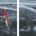
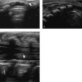
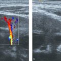
Examination of the pancreas with US usually occurs as part of an upper abdominal study, or of an upper and lower abdominal study. It is important to start the ultrasound examination with an evaluation of the pancreas before the child swallows air by talking or crying, and before air normally migrates toward the gastric antrum and transverse colon. The patient is scanned in a supine position. The hepatic left lobe is used as an appropriate window, combined with a subxiphoid transverse view. An oblique transverse view, following slight rotation counterclockwise parallel to the oblique orientation of the organ, evaluates as much of the body and tail as possible ( Fig. 12.1 ). More caudal views in the same direction, in an attempt to visualize the uncinate process of the pancreas, may be achieved by displacing the probe more caudally or by tilting the probe toward the patient’s feet ( Fig. 12.2 ). The distal body and tail of the pancreas may prove difficult to visualize anteriorly, and a rotated clockwise coronal view through the splenic hilum while the patient is turned toward the right ( Fig. 12.3 ), or through the left kidney while the patient is lying prone, may prove useful. Longitudinal views of the head and body through the hepatic left lobe complement the evaluation ( Fig. 12.4 ).

Fig. 12.1 Normal pancreas in a 4-year-old boy. The test was performed without preparation. The position of the probe rotated slightly counterclockwise, shown at the right lower part of the image, allowed a demonstration of a large part of the tail (t). h, head; i, isthmus; b, body; S, empty stomach. The echogenic linear structure coursing at the center of the organ represents the middle part of the pancreatic duct (arrow). Arrowheads indicate the pancreas.
Fig. 12.2 a, b a Echogenic normal neonatal pancreas (between arrowheads) seen during a subxiphoid transverse scan with minimal counterclockwise rotation. The liver (L) serves as an acoustic window. The area of the common bile duct in its intrapancreatic portion is minimally suggested (open arrow). Important landmarks include the confluence of the splenic vein and the portal vein (PV), the aorta (AO), and the inferior vena cava (IVC). Note that the gastric contents (S) may exhibit echogenicity similar to that of the neonatal pancreas. b Transverse scan with the same minimal counterclockwise rotation as in a, at a slightly more caudal level. The pancreatic head (h), lateral to the superior mesenteric vein (V), contains the distal common bile duct (small open arrow). Its posterior and medial part is the uncinate process (arrowhead). Important landmarks include the aorta (open arrowhead), inferior vena (white arrow), and superior mesenteric artery (A), etc. (A). S, gastric antrum; L, liver.
Fig. 12.3 Same neonate as in Fig. 12.2 . Longitudinal scan with and without color Doppler through the spleen (spl) visualizes the echogenic lateral aspect of the pancreatic tail (t) at the splenic hilum. Note the area of the splenic vein and artery (open arrows) and the neonatal left adrenal gland (arrowhead).
Fig. 12.4 a, b a Right parasagittal scan through the hepatic left lobe (L) in a 7-year-old girl. Part of the pancreatic head (h) is seen posterior to the stomach (S), inferior to the portal vein (PV), and anterior to the inferior vena cava (IVC). Note the hepatic artery (white arrowhead) and the right renal artery (open arrow), seen as dots because they run horizontally by the hepatic hilum and posteriorly to the IVC, respectively. b Left parasagittal scan through the liver left lobe (L) of the same patient. Part of the pancreatic body (b) is seen posterior to the liver and stomach (S), anterior to the obliquely coursing superior mesenteric vein (arrowhead), and anterior to the superior mesenteric artery (sma) coming off the aorta (AO). c, celiac trunk; arrow, left renal vein. The superior mesenteric vein is usually slitlike or ovoid at this section.

Gaseous distention of the stomach, small bowel, or colon may compromise an evaluation of the pancreas. Gentle or graded compression and patience are usually what works during sonography of the pancreas, together with tilting the probe toward the patient’s feet and slight rotation counterclockwise. Drinking liquids may not be as effective in children as in adults because children tend to swallow air as well, unless the examiner allows enough time for the bubbles to settle ( Fig. 12.5 ). One trick is to turn the patient on the right side for a few minutes and then back to a supine position, so that liquids may occupy the antrum and gas may move toward the fundus and colonic flexures for a few seconds, enough time to compress with the probe ( Fig. 12.6 ). The pancreas is better visualized after fasting; therefore, lastly, you may repeat the test following a 4-hour fast in children and a 3-hour fast in neonates.

Fig. 12.5 Transverse abdominal scan in a 7-year-old child. Liquids in the stomach (S) enable identification of all relevant to pancreatic ultrasound landmarks in order of importance: 1, aorta; 2, inferior vena cava; 3, superior mesenteric artery; 4, splenic vein; 4b, portal venous confluence; 5, gastric wall (arrowheads); D, duodenum; V, vertebra.
Fig. 12.6 a, b Ultrasound of an obese 11-year-old boy. a It is difficult to demonstrate the pancreas because of gas, which casts acoustic shadows and reverberations (g). b Following rotation of the patient to the right decubitus position and back in the supine position 4 minutes later, partial visualization of the pancreas (arrowheads) behind the stomach is feasible. The arrow points to the posterior wall of the antrum. The gas has moved back to the gastric fundus (g).


Tips from the Pro


  • The pancreas should be the first organ evaluated during an upper abdominal study and the second organ (after the urinary bladder) during an upper and lower abdominal US examination.


  • Scanning the child in the prone, lateral decubitus, or erect position may improve visualization through the liver or left kidney and may move the transverse colon out of the way. Asking a cooperative child to exhale or push the tummy out for seconds may also help.


12.2 Normal Anatomy, Variants, and Pseudo-lesions


The pancreas is a long, unencapsulated, retroperitoneal organ that has an oblique orientation. It extends from the duodenal loop medially to the splenic hilum laterally and is located in the anterior pararenal space at the upper part of the abdomen, anterior to the splenic vein and portal venous confluence ( Fig. 12.1 , Fig. 12.2 , Fig. 12.5 , Fig. 12.7 ). The pancreas is comma-shaped on transverse scans with a uniform echotexture and a smooth outline.

Fig. 12.7 Transverse section of the pancreas (white arrowheads) in a 6-year-old child showing the relationship of the pancreas to the surrounding structures and vascular landmarks. The pancreatic head (h) is near the duodenal loop (D) and wraps around the portal venous confluence (*). Note the hooklike uncinate process (open arrow). The superior mesenteric artery and surrounding echogenic fat form a doughnut posterior to the splenic vein (sv). 1, aorta; 2, inferior vena cava; 3, left renal vein; 4, left renal artery. The gastroduodenal artery, which runs anterior to the pancreatic head in a course similar to that of the common bile duct, is seen as an anechoic dot with a thin echogenic wall (open arrowhead). L, liver; LtK, left kidney; RtK, right kidney; S, stomach.

The pancreas consists of the head (which ends caudally and medially as the uncinate process), the isthmus (or neck), and the body and tail, with no clear distinction between them. The pancreatic head envelops the teardrop-shaped portal vein just at the junction of the splenic vein and superior mesenteric vein and extends caudally to wrap under the superior mesenteric vein as the uncinate process, which resembles a hook on transverse scans ( Fig. 12.2 and Fig. 12.7 ). This area is particularly important to examine because it includes the distal common bile duct and pancreatic duct and may reveal important pathology ( Fig. 12.8 ). A vascular landmark at this point is the gastroduodenal artery, which arises from the common hepatic artery and courses downward by the anterior surface of the pancreatic head or isthmus, parallel to the common bile duct ( Fig. 12.7 and Fig. 12.8 ).

Fig. 12.8 a, b One-month-old boy with jaundice and a history of prematurity. Ultrasound revealed areas of echogenicity in the gallbladder (not shown) following the administration of diuretics. a Three transverse sections though the pancreatic head, which was seen between the stomach (S) and the splenoportal junction (p) or at a slightly lower level between the stomach and superior mesenteric vein (v). The common bile duct is dilated (open arrowhead), and echogenicity representing choledocholithiasis (between cursors) is depicted in the two right panels. Note the gastroduodenal artery anteromedial to the pancreatic head (white arrowhead). Compare with the normal neonatal uncinate process in Fig. 12.12 . b Left: Longitudinal oblique section through the head of the pancreas (white arrowheads) shows dilatation of the common bile duct (open arrowhead) up to the choledochal stone (cursor). Right: Three days following medical treatment, the jaundice resolved and the common bile duct (open arrowhead) was normal. p, portal vein.

The neck or isthmus of the pancreas is the thinner part between the head and the body, anterior to the portal venous confluence ( Fig. 12.9 ). The pancreatic body lies posterior to the stomach or liver and anterior to the superior mesenteric artery. The superior mesenteric artery is anterior to the aorta and appears as the hole in a doughnut on transverse section because it is surrounded by a collar of echogenic fat ( Fig. 12.7 and Fig. 12.9 ). The bifurcation of the celiac artery into the hepatic and splenic arteries resembles a seagull in flight and is an important vascular landmark for the upper part of the pancreatic body ( Fig. 12.10 ). The tail of the pancreas is located anterior to the splenic vein and extends to the splenic hilum ( Fig. 12.1 and Fig. 12.3 ).

Fig. 12.9 Oblique longitudinal view with counterclockwise rotation pointing toward the patient’s right shoulder. The isthmus is visualized behind the pylorus and anterior to the splenoportal confluence. 1, splenic vein; 2, splenoportal confluence; 3, portal vein. Other visible landmarks include the gastric wall (between arrowheads), aorta (A), inferior vena cava (IVC), duodenal bulb (d), and superior mesenteric artery (4) with surrounding echogenic fat resembling a hole in a doughnut (open arrowheads).
Fig. 12.10 Transverse view of the cephalic pancreas visualizing the celiac trunk (C) and bifurcation. The common hepatic artery (white arrowheads) and the splenic artery (open arrowheads) resemble the open wings of a seagull. The pancreatic body (B) and tail (T) are visualized in association with this landmark if the probe is tilted downward. A, aorta.

Nonvascular landmarks are the pancreatic duct and common bile duct. The common bile duct courses downward parallel to the gastroduodenal artery and posterior to the pancreatic head, and it may not be visible in children unless dilated ( Fig. 12.8 and Fig. 12.11 ). The pancreatic duct courses centrally within the pancreatic parenchyma from the tail to the head, and parts of it can be identified by US as an echogenic line ( Fig. 12.1 ) or as an echo-free lumen with echogenic walls ( Fig. 12.12 ). A ductal diameter of 1.5 mm or more should be considered abnormal in children up to 6 years age until proved otherwise. The hypoechoic muscular wall of the stomach should not be mistaken for the pancreatic duct, especially when the body is imaged obliquely and the gastric contents are isoechoic to the pancreas and may be erroneously mistaken for the pancreatic body ( Fig. 12.13 ).

Fig. 12.11 a, b Visibility of the common bile duct during pediatric ultrasonography (US). a Transverse scan through the pancreatic head. b Corresponding magnetic resonance image, T2-weighted sequence. The normal terminal part of the common bile duct (arrow, a) is not visible or barely visible on US while it is a hyperintense dot (arrow, b) within the pancreatic head (h). Note how the various landmarks look with both methods. Ao, aorta; IVC, inferior vena cava; h, pancreatic head; u, uncinate process; PV, portal venous confluence; D, duodenal loop; g, gallbladder. The open arrows point toward the superior mesenteric artery. Note the visible diaphragmatic crus (arrowheads), occasionally seen at this level.
Fig. 12.12 Part of the main pancreatic duct of Wirsung is seen as an echo-free lumen with echogenic walls (between ‘x’s). Note the similarity to the alternating hyperechoic serosa, hypoechoic muscular layer, and hyperechoic submucosa of the gastric wall (arrowheads).
Fig. 12.13 a-d Examples of the gastric wall looking like a lucent tube with echogenic walls (open arrowheads). This structure simulates the pancreatic duct; however, it is located more anteriorly than the expected pancreatic duct and is adjacent to the gastric contents (g). Compare also with Fig. 12.12 . a Neonate. b Eight-year-old. c Four-month-old. d Sixteen-year-old.

The size of the pancreas varies depending on age. Measurements are shown in Fig. 12.14 and normal values in Table 12.1 . As a rule of thumb, when the thickness of the body is more than 1.5 cm, the pancreas is enlarged. The pancreatic head appears relatively voluminous compared with the body in children, which is normal. Care should be taken not to include the duodenal loop in the measurements or in the appreciation of the pancreatic head ( Fig. 12.15 ).


Table 12.1 Normal values of the pancreas in children

Age


Head


Body


Tail


Duct of Wirsung


Newborn infants


1.0 (0.4)


0.6 (0.2)


1.0 (0.4)


1 month–1 year


1.5 (0.5)


0.8 (0.3)


1.2 (0.4)


1–3 years


1.7 (0.3)


1.0 (0.2)


1.8 (0.4)


1.13 (0.15)


3–5 years


1.7 (0.3)


1.0 (0.2)


1.8 (0.4)


1.35 (0.15)


5–10 years


1.6 (0.4)


1.0 (0.3)


1.8 (0.4)


1.67 (0.17)


10–12 years


2.0 (0.5)


1.1 (0.3)


2.0 (0.4)


1.78 (0.17)


13–15 years


2.0 (0.5)


1.1 (0.3)


2.0 (0.4)


1.92 (0.18)


16–19 years


2.0 (0.5)


1.1 (0.3)


2.0 (0.4)


2.05 (0.15)


Abbreviation: SD, standard deviation. Source: Reprinted with permission of the Radiological Society of North America from Siegel MJ, Martin KW, Worthington JL. Normal and abnormal pancreas in children: US studies. Radiology 1987;165(1):15–18.


Note: In total, 273 patients (sex distribution not reported) were included in this retrospective ultrasound study. The maximum anteroposterior diameters of the head, body, and tail of the pancreas ( Fig. 12.14 ) were measured on transverse and oblique images.

Fig. 12.14 Anteroposterior diameter of the head (1) and body (2) and oblique anteroposterior diameter of the tail (3) in a 10-year-old child.
Fig. 12.15 a, b Scans of the pancreatic head a few seconds apart in a 6-month-old infant examined for gastroesophageal reflux. a There is an impression of a voluminous pancreatic head (h). b The duodenal loop (d) filled with fluid, and the true borders of the pancreatic head (arrowheads) and uncinate process (u) are appreciated.

The echogenicity of the pancreas varies; in general, it is equal to or slightly greater (but not much) than that of the normal liver, and its echotexture is smooth or minimally coarse ( Fig. 12.16 ). In about 10% of infants and children, it may be less echogenic than the liver. Care should be taken to evaluate the liver echogenicity as well, and not to mistake an isoechoic pancreas for a hypoechoic one in cases of hepatic steatosis ( Fig. 12.17 ) or for a hyperechoic one in cases of a hypoechoic liver due to hepatitis, in which the liver is also characterized by a starry sky appearance of the hepatic parenchyma.

Fig. 12.16 a–d Four examples of different degrees of echogenicity of the pancreas (p) in relation to the liver (L). The echotexture is smooth (a, d) or minimally coarse (b, c and Fig. 12.14 ). a Echogenic pancreas in a premature baby with a gestational age of 25 weeks. b Mildly echogenic in a 7-year-old. c Almost isoechoic in a 6-year-old. d Isoechoic in an 11-year-old.
Fig. 12.17 a, b False impression of a hypoechoic pancreas in a 4-year-old. a The pancreas (p) is hypoechoic relative to the liver (L). b Upon inspection of the liver (L), the echogenicity is typical of hepatic steatosis with focal fatty sparing (open arrow) by the gallbladder (g).

An important feature is the different echogenicity of the posterior pancreatic head and uncinate process. This area originates embryologically from the ventral anlage and exhibits a hypoechoic appearance because of its relatively low fat content. It has a geographic border like an area of focal fatty sparing and no mass effect ( Fig. 12.18 ). A cause of pseudo-lesions is the acoustic shadowing produced by the gastric contents ( Fig. 12.19 ) or by the fibrofatty ligamentum teres ( Fig. 12.20 ). Scanning with slight obliquity should help to distinguish a true focal lesion from a pseudo-lesion.

Fig. 12.18 a, b Hypoechoic appearance of the head and uncinate process in a 16-year-old girl. a Transverse scan shows a hypoechoic posterior pancreatic head and uncinate process (open arrowheads). b Longitudinal scan shows the hypoechoic derivatives of the ventral anlage (v) and the hyperechoic derivatives of the dorsal anlage in the remaining head (d). Note the lack of mass effect and lack of common bile duct (arrow) dilatation. Ao, aorta; IVC, inferior vena cava. See also embryology in Fig. 12.21 .
Fig. 12.19 a, b Pseudo-lesion due to gastric contents in a 2-year-old. a Shadowing (open arrowhead) over the pancreatic head gives the impression of a focal lesion (open arrow). b Oblique scanning of the same region confirms a normal pancreatic head (open arrowheads).
Fig. 12.20 a, b Pseudo-lesion in a 6-year-old. a Shadowing over the pancreatic head behind the ligamentum teres (open arrowhead) gives the impression of a hypoechoic lesion (open arrow). b Oblique scanning of the same region confirms a normal pancreatic head (open arrowheads).


Tips from the Pro


  • Knowledge of the vascular and nonvascular landmarks and multiple views with and without obliquity ensure visualization of all the pancreatic parts and avoid the misinterpretation of pseudo-lesions as true lesions. Routine measurements of the pancreas and pancreatic duct, and appreciation of its echogenicity compared with that of the liver, may increase the sensitivity of US in the detection of focal and diffuse pancreatic abnormalities.


12.3 Pathology


12.3.1 Developmental Anomalies


A knowledge of basic embryology is helpful in understanding the morphology and clinical importance of the most common developmental pancreatic abnormalities ( Fig. 12.21 ). The ventral anlage forms the uncinate process and part of the pancreatic head, while the dorsal anlage forms the remaining, larger part of the organ.

Fig. 12.21 a–d Schematic representation of normal development and the more common congenital abnormalities of the pancreas. a The ventral and the dorsal pancreas have formed off the primitive gut from the ventral and dorsal buds, respectively. The ventral segment rotates around and posterior to the duodenum, together with the distal biliary tree. b The ventral and dorsal pancreas fuse. At this point, the pancreatic ducts do not communicate. In case development stops, this configuration corresponds to pancreas divisum. c Finally, a wide communication between the two ducts occurs. The duct of the ventral pancreas becomes the duct of Wirsung and drains into the major papilla together with the common bile duct. The duct of the dorsal pancreas at its distal part becomes the main pancreatic duct or part of the duct of Wirsung. At its proximal part it involutes, becoming smaller or atretic, and is then called the accessory pancreatic duct of Santorini, which drains into the minor papilla when intact. This is the normal completed evolution of the pancreas. d Incomplete rotation of the ventral anlage results in a tongue of pancreatic tissue surrounding the duodenum. The pancreatic duct may be seen encircling the duodenum.

Pancreas divisum is the most common congenital anomaly of the pancreatic ductal system and is encountered in 4 to 10% of the population. Pancreas divisum results from failure of fusion between the ventral and dorsal pancreatic ducts ( Fig. 12.21 c). The ventral duct (duct of Wirsung) drains only the ventral pancreatic anlage, whereas most of the gland empties into the minor papilla through the dorsal duct (duct of Santorini). Pancreas divisum may be asymptomatic or present with chronic abdominal pain and recurrent pancreatitis in children ages 5 to 15 years because of functional stenosis at the minor papilla. US usually fails to diagnose this condition. Endoscopic retrograde pancreatography or magnetic resonance (MR) pancreatography demonstrates noncommunicating dorsal and ventral ducts, independent drainage sites, and a dominant dorsal pancreatic duct. The ventral duct is typically short and narrow.


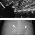
Annular pancreas is a rare congenital anomaly (1 in 2,000 persons) in which incomplete rotation of the ventral anlage causes a segment of the pancreas to encircle the second part of the duodenum ( Fig. 12.21 d). It occurs either in isolation or together with other congenital abnormalities, including tracheoesophageal fistula, duodenal stenosis or atresia in infants, Down syndrome, and congenital heart defects. Annular pancreas presents with proximal obstruction causing a “double-bubble” sign on radiographs in neonates, and with bile duct obstruction, associated pancreatitis, and “peptic ulcer”-like disease in older children. Annular pancreas can be diagnosed by US as pancreatic tissue encircling the duodenum, with proximal duodenal dilatation ( Fig. 12.22 ). Computed tomography (CT) and MR imaging may confirm these findings. Magnetic resonance cholangiopancreatography (MRCP) may demonstrate an annular duct encircling the descending duodenum.

Fig. 12.22 a–d Annular pancreas. a There is lateral extension of the pancreatic head (p) with a triangular tongue of tissue (arrowheads) anterior to a dilated proximal duodenum (arrow) and a collapsed descending duodenum, which is a hypoechoic structure with the gut signature (*). The anterior arrow indicates the gastric wall. b The descending duodenum is seen with a collapsed lumen (arrowheads) coursing at the center of the pancreatic head. The arrow indicates the anterior surface of the pancreatic head. The open arrowheads indicate lateral surface of the pancreatic head encircling the duodenum. c Magnetic resonance imaging, axial true FISP (fast imaging with steady-state precession) sequence, shows the stomach and duodenum fluid-filled (arrows). Pancreatic tissue (*) almost completely encircles the duodenum (arrowhead). d Axial T2-weighted sequence lower confirms that the duodenum (arrow) enters the center of the pancreatic head and contains fluid and kissing folds (arrowhead). (Case courtesy of Dr. Lil-Sofie Ording Müller, Norway.)

Ectopic pancreas occurs in 0.6 to 13.7% of the population and may be found in the stomach, duodenum, jejunum, a Meckel diverticulum, or ileum, and rarely in other abdominal sites ( Fig. 12.23 ). The ectopic tissue measures around 0.5 to 2.0 cm in diameter and is usually located in the submucosa. Ectopic pancreas is asymptomatic, although complications due to mass effect (stenosis, intussusception), ulceration, bleeding, pancreatitis, cystic degeneration, or malignancy may develop.

Fig. 12.23 a, b Ectopic pancreas. a Gastric pancreas. There is a mural lesion with the characteristic smooth echotexture of normal pancreas (p). Note the intact overlying mucosa, seen as a thin, hyperechoic layer (arrowheads). L, fluid within the gastric lumen; C, pyloric channel with hypoechoic muscular layer (open arrowheads). b Ectopic pancreas in a Meckel diverticulum, seen as a nodule with a characteristic echotexture (between cursors) at the periphery of the diverticulum (arrows). The echogenic luminal contents (*) are due to a previous barium study. (Case courtesy of Dr. Paolo Toma, Italy.)

Other congenital abnormalities include pancreatic agenesis, which is extremely rare and incompatible with life, and partial agenesis or hypoplasia or congenitally short pancreas, which can occur in isolation or be associated with heterotaxy/polysplenia syndromes. The pancreas in hypoplasia consists of a short, enlarged, or globular pancreatic head and agenesis of the pancreatic body and tail due to the absence of dorsal anlage development ( Fig. 12.24 ). Patients who have agenesis of the dorsal pancreas often present with nonspecific abdominal pain, which may or may not be caused by pancreatitis, and they are at increased risk for diabetes mellitus. Situs inversus is inversion of the intra-abdominal contents and anatomical landmarks, with an intact pancreas ( Fig. 12.25 ).

Fig. 12.24 a–f Heterotaxy syndrome with congenitally short pancreas, polysplenia, malrotation, preduodenal portal vein, biliary atresia, and azygos continuation. a Transverse scan shows a globular hourglass-shaped pancreas (outlined by arrowheads) between the superior mesenteric artery (a), stomach (S), and portal vein (p). b At a more caudal level, the remaining lower head and uncinate process (arrowheads) are seen around the splenoportal confluence (psv). c At a more caudal level, the superior mesenteric vein (v) is located anterior and to the left of the superior mesenteric artery (open arrow), consistent with malrotation. The portal vein (p) is seen anterior to the duodenum (d; s, stomach). There is no pancreatic body or tail visible. d Longitudinal scan shows the portal vein (p) in gray-scale and color Doppler imaging lying anterior to the duodenum (open arrow), consistent with a preduodenal portal vein. e Multiple splenules (s) are identified on the left. f Computed tomographic evaluation. A nearly symmetric lobulated liver extending to the left hypochondrium (L) is associated with a midline gallbladder (g) and a portal vein (open arrows) anterior to the duodenum (white arrowheads). Note the polysplenia (*), hypoplastic or congenitally short pancreas (p), and absence of the dorsal pancreas. The left renal vein (v) drains into the azygos continuation (open arrowheads), which is best demonstrated on coronal reconstructions (bottom left). S, stomach. (Case courtesy of Dr. I. Gassner, Austria.)
Fig. 12.25 a–c Situs inversus. Transverse section of the pancreas (a, b) and coronal T2-weighted magnetic resonance (MR) imaging (c) in an 18-month-old boy investigated for neuroblastoma. The presence of situs inversus is easily diagnosed by the position of the large, solid abdominal organs and also by the inverted position of major vascular landmarks such as 1, aorta; 2, inferior vena cava; 3, superior mesenteric artery; and 4, portal venous confluence. MR imaging confirms the left-sided position of the pancreatic head (h), a right-sided tail (t), and inversion of the liver (L) and spleen (S).

Only gold members can continue reading. Log In or Register to continue

Stay updated, free articles. Join our Telegram channel

Jun 9, 2020 | Posted by in ULTRASONOGRAPHY | Comments Off on 12 Pancreas

Full access? Get Clinical Tree
Get Clinical Tree app for offline access