Clinical Presentation
A 13-month-old female infant presents with swelling of the left arm.
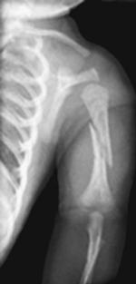
Figure 120A
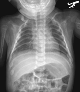
Figure 120B
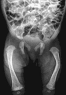
Figure 120C
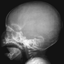
Figure 120D
Radiologic Findings
Frontal radiograph of the left humerus (Fig. 120A) shows an oblique fracture of the mid-shaft of the left humerus. Frontal view chest radiograph (Fig. 120B) demonstrates osteopenia and gracile ribs with multiple healing fractures. A fracture of the left humerus is also visualized. Radiograph of the lower extremities (Fig. 120C) demonstrates osteopenia and bowing of the femurs, greater on the left. Lateral view of the skull (Fig. 120D) demonstrates multiple wormian bones.
Diagnosis
Osteogenesis imperfecta (OI)
Differential Diagnosis
- Nonaccidental injury/child abuse
- Menkes (kinky hair) syndrome
- Hypophosphatasia
- Campomelic dysplasia
Discussion
Background
OI is one of the most common genetic disorders of connective tissue. OI is characterized by the following features: fragile bones, frequent fractures, limb bowing, spinal shortening, blue sclerae, deafness, poor dentition, ligamentous laxity, and easy bruising. Approximately 4 in 100,000 births present with OI.
Etiology
OI results from an abnormal quantity and/or quality of type I collagen. Mutations in the COL1A1 gene on chromosome 17 or COL1A2gene on chromosome 7 alter the structure of the triple helix of collagen. This results in decreased synthesis of collagen or structurally abnormal collagen. Abnormalities in the collagen type I and collagen type III are detected in fibroblast cultures from skin extracts of patients with OI.
Clinical Findings
Variability in clinical features has led to the concept of heterogeneity based on a classification of four major types of OI, as shown in Table 120A.
Complications
- Nonunion of fractures
- Flail chest
- Osteomyelitis
- Osteosarcoma (rare)
- Basilar impression with brain stem compression
- Cardiomyopathy, mitral, or aortic valve disease
Pathology
Stay updated, free articles. Join our Telegram channel

Full access? Get Clinical Tree


