CASE 123 A 63-year-old man presents with epigastric distress and a history of recent cholecystectomy (Fig. 123.1). Fig. 123.1 Fig. 123.2 • The patient should take nothing by mouth between less than 24 hours and more than 4 hours before the test. • Immediately following the injection of 1.5 mCi of 99mTc-DISIDA, the patient is placed in a supine position beneath the gamma camera. • Continuous images acquired in a 64 × 64 matrix are divided into 1-minute frames. • Images are obtained in the left anterior oblique projection with a slant-hole collimator to better separate the small bowel from the liver. Straight anterior views may be obtained with a high-resolution, parallel-hole collimator if a slant-hole collimator is not available. • Delayed static images may be acquired as necessary. Selected 1-minute images (Fig. 123.1
Clinical Presentation
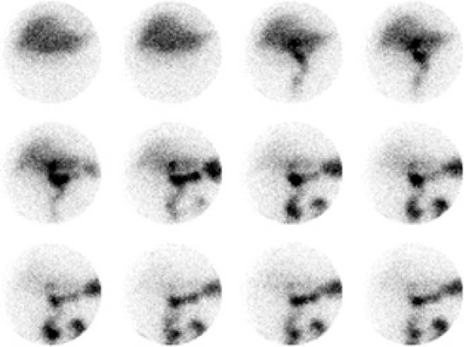
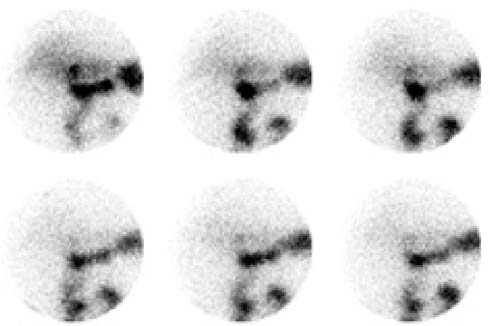
Technique
Image Interpretation
![]()
Stay updated, free articles. Join our Telegram channel


Full access? Get Clinical Tree


