CASE 127 A 53-year-old woman who has hepatitis C presents with right upper quadrant pain associated with meals (Figs. 127.1 and 127.2). Fig. 127.1 Fig. 127.2 • The patient should take nothing by mouth between less than 24 hours and more than 4 hours before the test. • Immediately following the injection of 5 mCi of 99mTc-DISIDA, the patient is placed in a supine position beneath the gamma camera. • Continuous images acquired in a 256 × 256 matrix for 1 hour are divided into 1-minute frames. • Images are obtained with a high-resolution, parallel-hole collimator. • Following the collection of images for 1 hour, sincalide (0.02 μg/kg) is administered intravenously over 60 minutes while a second series of 1-minute images is obtained. Selected 1-minute images (Fig. 127.1) immediately after the injection of tracer demonstrate normal uptake of tracer in the liver and subsequent excretion into the gallbladder. The gallbladder contour is slightly irregular, which may be secondary to a fold, or “Phrygian cap.”
Clinical Presentation
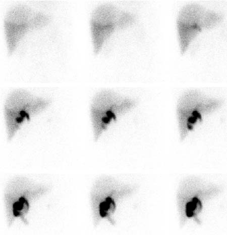
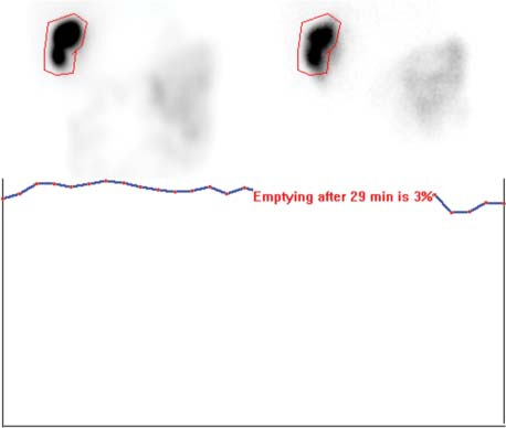
Technique
Image Interpretation
Related posts:


Stay updated, free articles. Join our Telegram channel

Full access? Get Clinical Tree


