Case 13
 Clinical Presentation
A 39-year-old woman with left flank pain. Computed tomography urinary stone protocol was performed.
 Imaging Findings
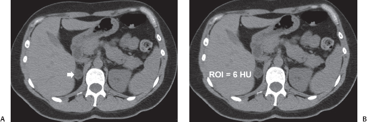
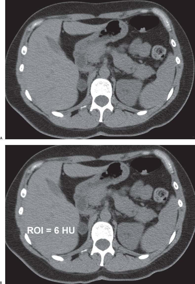
(A) Noncontrast computed tomography (CT) image at the level of the adrenal glands shows a nodule in the expected location of the right adrenal gland (arrow). The nodule is oval, well defined, and homogeneous. It does not show calcifications or areas of macroscopic fat. (B) Attenuation has been measured within the nodule. The region of interest is of adequate size and satisfactorily placed. The attenuation reading within the region of interest is 6 Hounsfield units (HU).
 Differential Diagnosis
• Adrenal cortical adenoma: On noncontrast CT, a well-defined lesion that has no wall, no macroscopic fat, and homogeneous attenuation with a value of < 10 HU is characteristic of adrenal cortical adenoma.
• Adrenal cyst:
Stay updated, free articles. Join our Telegram channel

Full access? Get Clinical Tree


