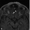
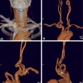
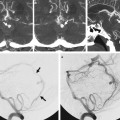
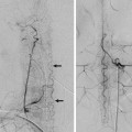
Fig. 13.1 Unenhanced CT (a) demonstrates a right apical frontoparietal hemorrhage. CTA (coronal reformats: b,c,d; 3D reconstruction: e) revealed, as the source for the hemorrhage, an AVM fed by the distal ACA (inferior parietal branch) with a focal intranidal outpouching (arrow) that was held responsible for the bleed. Drainage of this monocompartimental AVM was directed into a median parietal vein. Case is continued in ▶ Fig. 13.2.
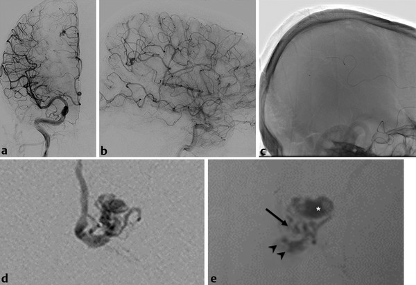
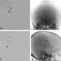
Fig. 13.2 (a–e) Angiography with right internal carotid artery injection (anteroposterior and lateral) confirmed the diagnosis. A microcatheter was advanced into the distal inferior parietal branch (c), and the subsequent microcatheter injection (d) demonstrated the AVM, the intranidal aneurysm, and the drainage. The glue cast after liquid embolic embolization demonstrates occlusion of the intranidal aneurysm (asterisk), the distal feeding artery (arrow), and the proximal draining vein (arrowheads). Case is continued in ▶ Fig. 13.3.
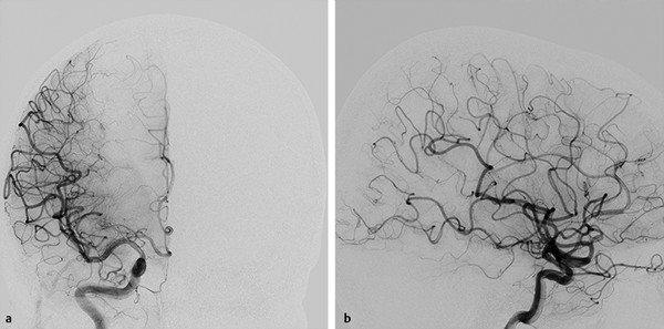
Fig. 13.3 Angiography in anteroposterior and lateral views (a,b) after embolization reveals complete occlusion of the AVM.
13.1.3 Diagnosis
Ruptured arteriovenous malformation (AVM) fed by the inferior parietal branch of the distal anterior cerebral artery (ACA).
13.2 Embryology and Anatomy
Similar to the anatomy of the cortical branches of the middle cerebral artery (MCA), which are discussed in Case ▶ 16
Stay updated, free articles. Join our Telegram channel

Full access? Get Clinical Tree


