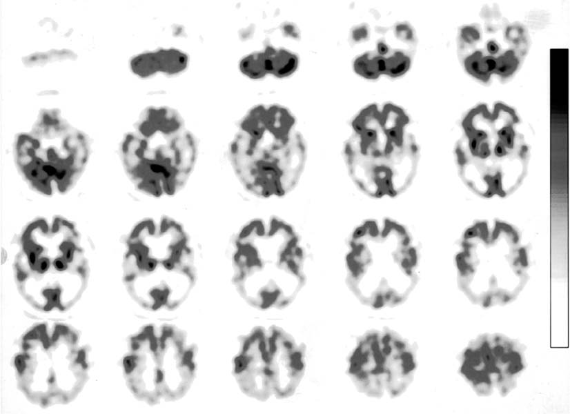
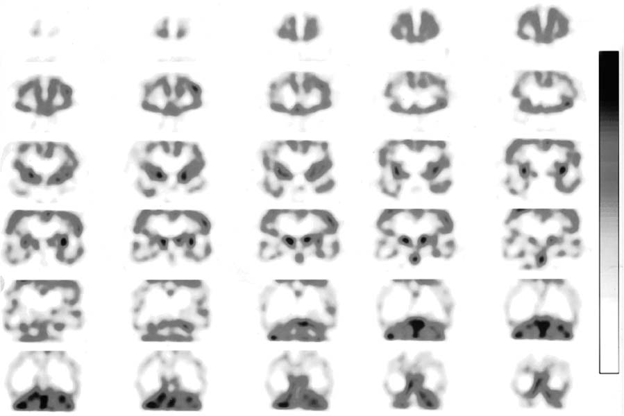
CASE 132 A 62-year-old woman presents with short-term memory loss that has increased over the past 3 years. A radionuclide brain perfusion SPECT study is ordered to evaluate the perfusion pattern. Fig. 132.1 Fig. 132.2 • Approximately 20 mCi of 99mTc-HMPAO or 99mTc-ECD is injected intravenously. • Imaging time is 20 to 30 minutes after injection (1 hour if 99mTc-ECD is used). • Acquisition protocol is 30 minutes with a 360-degree SPECT rotation. • The patient should be supine, with the head slightly elevated and eyes closed. The head should be as close to the camera as possible and strapped tightly with a nonattenuating material such as Velcro, rubber, or elastic to avoid head motion. • During injection and image acquisition, the room should be quiet, with the lights dimmed. • The head should be in the center of the axis of rotation. The axial (Fig. 132.1) and coronal (Fig. 132.2) views display a global reduction in radiotracer uptake with increased background activity. Lateral frontal perfusion defects are seen on the left side. Marked reduction in the perfusion to the temporal lobes (medial, lateral, and inferior aspects) is seen bilaterally in the axial and coronal slices. The left side is more affected than the right side. Marked reduction in the perfusion to the entire parietal cortex is noted bilaterally. Symmetric radiotracer uptake is visible in the basal ganglia and thalami. • Alzheimer disease
Clinical Presentation
Technique
Image Interpretation
Differential Diagnosis
Stay updated, free articles. Join our Telegram channel

Full access? Get Clinical Tree


