CASE 133
Clinical Presentation
A 34-year-old woman who has had intractable complex partial seizures since the age of 22 years and has undergone a prior left temporal lobectomy is admitted for evaluation of the seizure focus. Depth electrode placement shows spike activity in the frontal and temporal lobes.
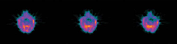
Fig. 133.1 Ictal Imaging.
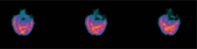
Fig. 133.2 Interictal imaging.
Technique
• Approximately 20 mCi of 99mTc-HMPAO or 99mTc-ECD is injected intravenously.
• Imaging time is 20 to 30 minutes after injection (1 hour if 99mTc-ECD is used).
• Acquisition protocol is 30 minutes with a 360-degree SPECT rotation.
• The patient should be supine, with the head slightly elevated and eyes closed. The head should be as close to the camera as possible and strapped tightly with a nonattenuating material such as Velcro, rubber, or elastic to avoid head motion.
• During injection and image acquisition, the room should be quiet, with the lights dimmed (avoid disturbances and noise).
• The head should be in the center of the axis of rotation.


