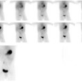
CASE 139 A 54-year-old woman presents with dysphagia. Fig. 139.1 • A 0.3 mCi dose of 99mTc-DTPA is mixed with cornflakes and milk. • Posterior view imaging begins just after ingestion of the meal. • Images are acquired at 10 seconds per frame for 30 minutes. • A region-of-interest is drawn on the computer for the esophagus. • A time–activity curve is generated. • Esophageal retention is quantified. Fig. 139.2 Sequential posterior images (Fig. 139.1) demonstrate delayed clearance from the esophagus, with a minimal amount of radiolabeled meal reaching the stomach. A region-of-interest is drawn around the esophagus (Fig. 139.2
Clinical Presentation
Technique
Image Interpretation
![]()
Stay updated, free articles. Join our Telegram channel

Full access? Get Clinical Tree


