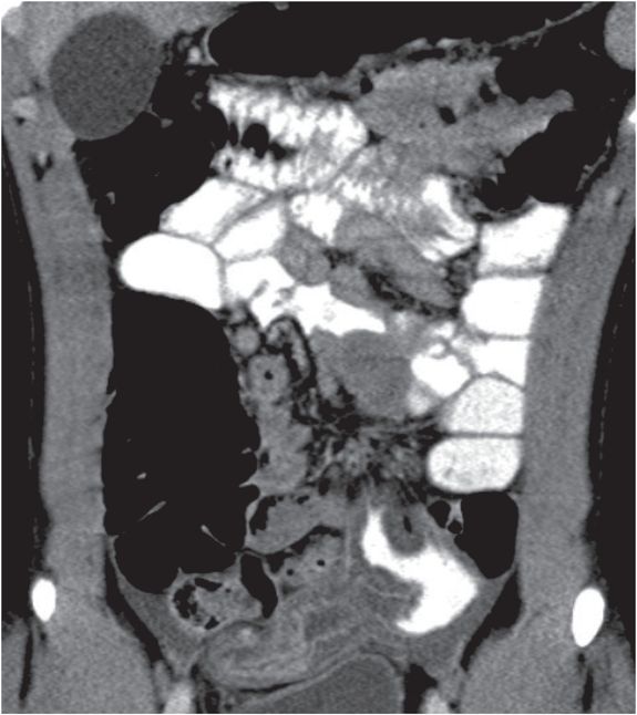
Diagnosis: Meckel’s diverticulitis
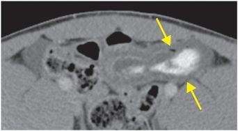
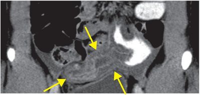
Axial (left image) and coronal (right image) contrast-enhanced CT images show a large, blind-ending tubular structure to the left of midline with thickened walls (arrows). There is some enteric contrast material within this structure, as seen on the axial image.
Discussion
Overview of Meckel’s diverticulitis
Meckel’s diverticulitis is acute inflammation of a Meckel’s diverticulum.
Meckel’s diverticulum is seen in 2% of the population and is the most common congenital gastrointestinal anomaly.
A Meckel’s diverticulum is the result of incomplete regression of the omphalomesenteric (vitelline) duct, which connects the intestine to the yolk sac via the umbilicus in early embryological life. There is a spectrum of anomalies due to incomplete atrophy of the omphalomesenteric duct, with the most rare and dramatic being an umbilicoileal fistula due to complete patency of the duct, which causes fecal drainage from the umbilicus in the neonatal period.
Meckel’s diverticulum is found on the antimesenteric aspect of the distal ileum, usually within 100 cm of the ileocecal valve.
Although there are many exceptions in real life, the “rule of twos” is often taught: Meckel’s are present in 2% of the population, are 2 feet from the ileocecal valve, usually 2 inches long, and usually become symptomatic before age 2.
Complications of Meckel’s diverticulum
While most Meckel’s diverticula are asymptomatic, between 4% and 40% of patients will experience a complication during their lifetime, including gastrointestinal hemorrhage, bowel obstruction, or diverticulitis (as in this case).
There can be ectopic gastric or pancreatic mucosa within the Meckel’s diverticulum, which predisposes to complications. Most cases of symptomatic Meckel’s diverticulitis occur in the pediatric population, where gastrointestinal hemorrhage is by far the most common complication.
A Meckel’s diverticulum may invaginate or invert into the small intestinal lumen, thereby serving as a lead point for ileocolic intussusception and small bowel obstruction (inverted Meckel’s diverticulum).
Stay updated, free articles. Join our Telegram channel

Full access? Get Clinical Tree


