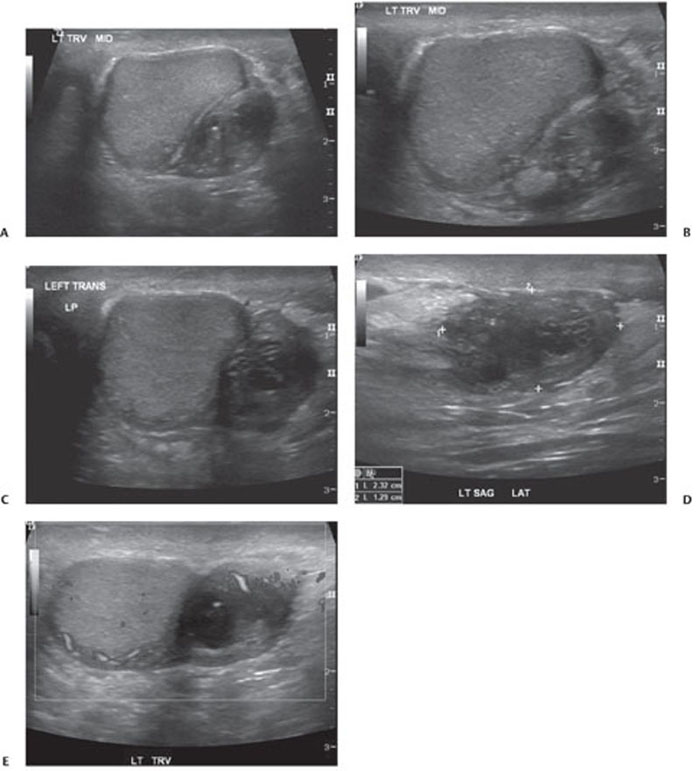
CASE 146 A 33-year-old man complains of a 2-month history of a nontender left testicular mass. Fig. 146.1 (A–E) Ultrasound images of the left scrotum show a normal-appearing left testis with a heterogeneous, predominantly hypoechoic left extratesticular mass with some Doppler flow. Ultrasound images of the left scrotum show a normal-appearing left testis with a heterogeneous, predominantly hypoechoic left extratesticular mass with some Doppler flow (Fig. 146.1). Left extratesticular adenomatoid tumor
Clinical Presentation
Radiologic Findings
Diagnosis
Differential Diagnosis
Related posts:


Stay updated, free articles. Join our Telegram channel

Full access? Get Clinical Tree


