CASE 65 A 78-year-old diabetic man presents with a spiking fever and flank pain. Fig. 65.1 Axial contrast-enhanced CT image in a diabetic patient with a history of urinary tract infection shows an ill-defined, hypodense corticomedullary renal abscess. Axial contrast-enhanced computed tomography (CT) image shows an ill-defined, hypodense left renal lesion with thick walls (Fig. 65.1). Renal abscess A renal abscess is an uncommon renal condition. It can be cortical or corticomedullary, depending on the route of infection. When left untreated, a renal abscess can lead to significant mortality and morbidity. With the use of image-guided intervention techniques and antibiotics, mortality and morbidity due to renal abscess are significantly reduced.
Clinical Presentation
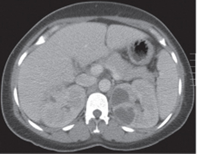
Radiologic Findings
Diagnosis
Differential Diagnosis
Discussion
Background
Clinical Findings
Complications
Related posts:


Stay updated, free articles. Join our Telegram channel

Full access? Get Clinical Tree


