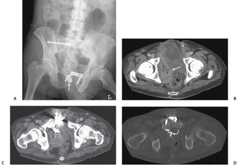
CASE 100 A middle-aged man with a recent history of a high-speed motor vehicle accident who underwent emergent open reduction and internal fixation of pelvic fractures presents months later with pelvic pain and fevers. Fig. 100.1 (A) Supine abdominal radiograph shows screw and plate fixations of the patient’s pelvic fractures. Partially obscured by the orthopedic hardware is a radiodense ribbon at the level of the pubis (arrow). (B–D) Contrast-enhanced CT images show a right pelvic abscess collection (arrow) in the right obturator space that demonstrates an enhancing rind around a spongiform gas collection with mass effect on the rectum. (D) The radiodense ribbon is best seen on bone windows and is within the collection (curved arrow). Supine abdominal radiograph shows screw and plate fixations of the patient’s pelvic fractures (Fig. 100.1A). Partially obscured by the orthopedic hardware is a radiodense ribbon at the level of the pubis. Contrast-enhanced computed tomography (CT) images show a right pelvic abscess collection in the right obturator space that demonstrates an enhancing rind around a spongiform gas collection with mass effect on the rectum. The radiodense ribbon is best seen on bone windows and is within the collection (Fig. 100.1B–D). Gossypiboma (textiloma or retained surgical sponge) Abscess as a complication of
Clinical Presentation
Radiologic Findings
Diagnosis
Differential Diagnosis
Related posts:


Stay updated, free articles. Join our Telegram channel

Full access? Get Clinical Tree


