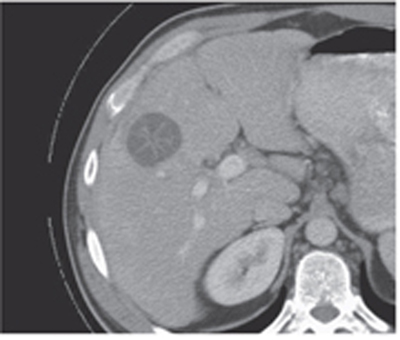
CASE 4 A 46-year-old man presents with right upper quadrant pain and tenderness. Fig. 4.1 An axial nonenhanced CT image of the liver shows a well-defined, round-shaped, hypodense lesion in the right lobe with hyperdense internal septa demonstrating a spoke-wheel pattern. Axial contrast-enhanced computed tomography (CT) image (Fig. 4.1) shows a hypodense, well-defined, round cystic lesion in the liver; thin, mildly enhancing internal septations arranged in a spoke-wheel pattern within the lesion are noted. Hydatid disease Hydatid disease is a worldwide parasitic infestation caused by different species of a tapeworm belonging to the genus Echinococcus; this zoonosis, quite uncommon in the United States and northern Europe, is otherwise widely present in the Mediterranean areas, the Middle East, the southern part of South America, Iceland, Africa, Australia, and New Zealand. Hydatid disease occurs in both sexes usually in their 3rd to 5th decade and affects the liver (75%), the lungs (15%), and, less commonly, the brain, kidney, spleen, and bone. Unusual locations (< 1%) include the gastric wall, orbit, heart, mediastinum, and adrenal glands. Hydatid cysts have a unique structure that can almost always be easily recognized at cross-sectional imaging. They present three different layers: the outer pericyst, the middle laminated layer, and the inner germinative membrane. Small, round vesicles (daughter vesicles) containing protoscolices are found within the mother cyst firmly attached to the germinative membrane. Clinical presentation mostly relies on parasite load, size, and location of the hydatid cysts. Some patients can be completely asymptomatic despite the presence of these lesions; other patients can present with abdominal pain and tenderness, fever, and jaundice due to the mass effect of the cysts in the liver.
Clinical Presentation
Radiologic Findings
Diagnosis
Differential Diagnosis
Discussion
Background
Clinical Findings
Complications
Related posts:


Stay updated, free articles. Join our Telegram channel

Full access? Get Clinical Tree


