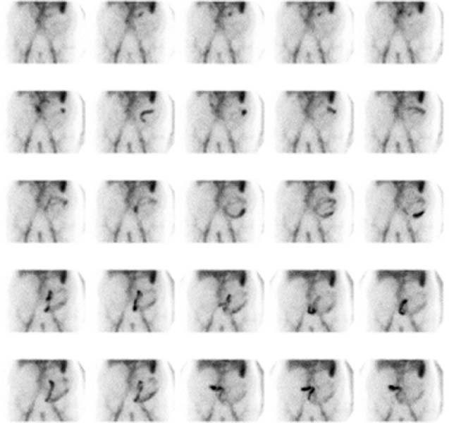
CASE 149 A 68-year-old man presents with intermittent melena and a recently falling hematocrit. Fig. 149.1 • Three milliliters of autologous red blood cells are labeled with 99mTc-pertechnetate (25 mCi) and injected intravenously. • The patient is placed in a supine position beneath a gamma camera with a large field of view. • Flow images are acquired at 1 to 3 seconds per frame for 60 seconds (not shown). • Anterior abdominal image data are acquired continuously (1 minute per frame for 90 minutes). • Delayed dynamic images (1 minute per frame for 30 minutes) are acquired as needed. Serial 1-minute anterior images over the abdomen (Fig. 149.1
Clinical Presentation
Technique
Image Interpretation
![]()
Stay updated, free articles. Join our Telegram channel


Full access? Get Clinical Tree


