Case 15

 Clinical Presentation
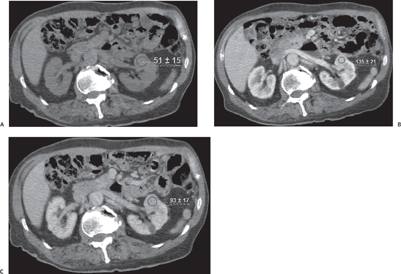
A 38-year-old man who underwent multiphase contrast-enhanced computed tomography to evaluate a focal renal lesion seen on noncontrast stone protocol computed tomography.
 Imaging Findings

(A) Computed tomography (CT) image at the level of the kidney before injection of intravenous (IV) contrast shows a contour deformity in the left kidney (arrowhead) due to a parenchymal mass with an attenuation value of 51 Hounsfield units (HU). (B) CT image at the same level obtained during the corticomedullary phase after injection of IV contrast shows enhancement of the mass to 135 HU. The enhancement is homogeneous. The visualized portion of the left renal vein (arrowhead) is normal. (C) CT image at the same level obtained during the nephrographic phase after injection of IV contrast shows some washout of the lesion with a decrease in attenuation to 93 HU. An additional small, simple cyst (arrow) has become apparent in the posterior portion of the left kidney. No retroperitoneal lymphadenopathy is seen in the images provided.
 Differential Diagnosis
• Renal cell carcinoma (RCC):
Stay updated, free articles. Join our Telegram channel

Full access? Get Clinical Tree


