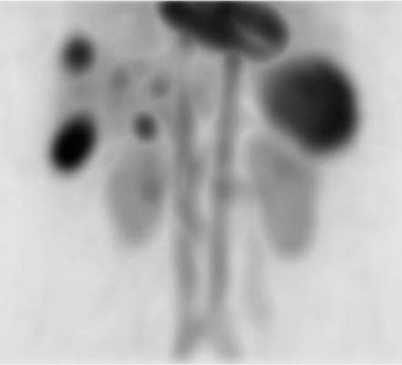
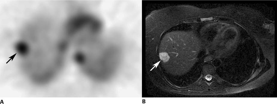
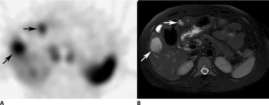
CASE 151 A 44-year-old woman undergoes abdominal ultrasonography, which reveals multiple echogenic lesions within the liver. This leads to a nuclear medicine tagged red blood cell (RBC) study (Figs. 151.1, 151.2, and 151.3). Fig. 151.1 Fig. 151.2 Fig. 151.3 • The in vitro or modified in vivo method is used to label autologous blood with 25 to 30 mCi of 99mTc-pertechnetate. • The patient is placed in a supine position and imaged anteriorly with a gamma camera that has a large field of view. The gamma camera is placed over the abdomen. • Initial flow images are acquired at 1 to 3 seconds per frame for 1 minute (not shown). • Anterior abdominal 1-minute images are acquired continuously for 60 to 90 minutes. Anterior maximum-intensity projection (MIP) view from the SPECT acquisition (Fig. 151.1
Clinical Presentation
Technique
Image Interpretation
![]()
Stay updated, free articles. Join our Telegram channel


Full access? Get Clinical Tree


