Case 16

 Clinical Presentation
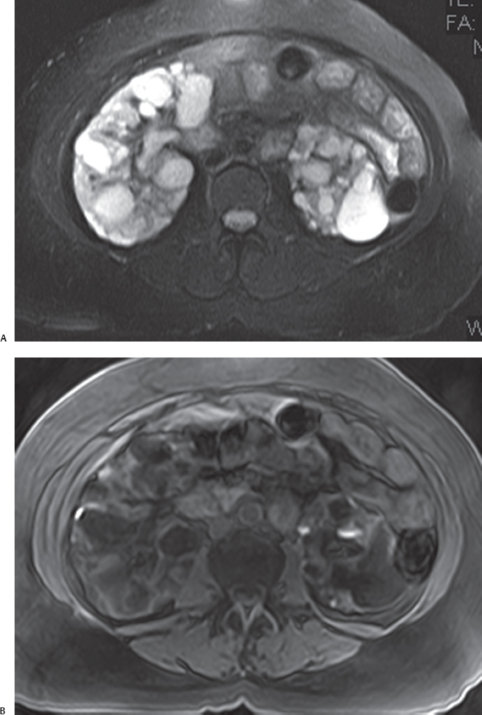
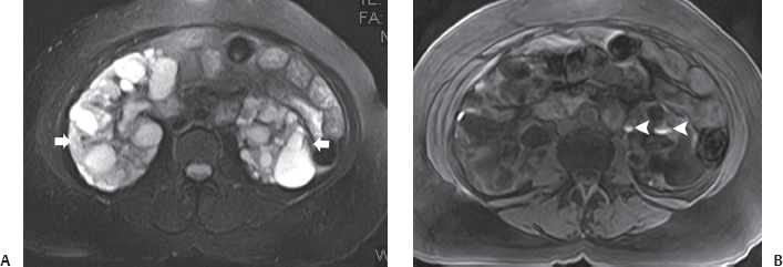
A 45-year-old woman with recently diagnosed renal insufficiency and palpable bilateral flank masses. Magnetic resonance imaging was performed for morphologic evaluation of the kidneys.
 Imaging Findings

(A) Axial fat-saturated T2-weighted magnetic resonance imaging (MRI) at the level of the kidneys shows that both kidneys are enlarged. The renal parenchyma on both sides has been replaced by multiple focal cystic lesions (arrows). The signal intensity of the cysts is variable, ranging from dark to very bright. (B) Fat-saturated T1-weighted MRI at the same level as Figure A shows the cysts to be of low signal intensity. Some of the cysts are hyperintense (arrowheads) because of recent hemorrhage.
 Differential Diagnosis
• Adult polycystic kidney disease (PKD): Numerous, tightly packed renal cysts that involve both kidneys and replace the renal parenchyma are typical of adult PKD. Most of the cysts are typical simple cysts. However, they have a propensity to hemorrhage, and the signal intensity may be variable depending on the amount and chronicity of hemorrhage.
• Multiple bilateral simple cysts:
Stay updated, free articles. Join our Telegram channel

Full access? Get Clinical Tree


