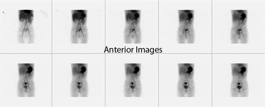
CASE 160 A 15-month-old child presents with the passage of bright red blood per rectum for 1 day and no abdominal pain or vomiting. Physical examination shows a pale infant with otherwise normal examination findings. Laboratory evaluation shows a hemoglobin level of 8 mg/dL. A bleeding Meckel diverticulum is considered as a possible diagnosis, and a Meckel scan is ordered. Fig. 160.1 • 99mTc-pertechnetate, 100 μCi (3.7 MBq) per kilogram. The maximum dose is 10 mCi (370 MBq), and the minimum dose is 200 μCi (7.5 MBq). • Ultra-high-resolution collimator, matrix 128 × 128. The acquisition of 1 frame per second for 60 seconds is followed by the acquisition of 1 frame per minute for 30 minutes. Static image matrix 256 × 256 for 60 seconds. • Nothing by mouth for 4 hours before the study. Images are taken anteriorly and posteriorly. The Meckel scan shows an abnormal focus of uptake in the lower midabdomen, just above the urinary bladder (Fig. 160.1). There is normal uptake in the stomach and normal excretion of tracer in the uri-nary bladder. • Enteric duplication cysts • Vascular malformations • Gastrogenic cyst • Inflammatory bowel disease
Clinical Presentation
Technique
Meckel Scan
Image Interpretation
Differential Diagnosis
Diagnosis and Clinical Follow-Up
Related posts:


Stay updated, free articles. Join our Telegram channel

Full access? Get Clinical Tree


