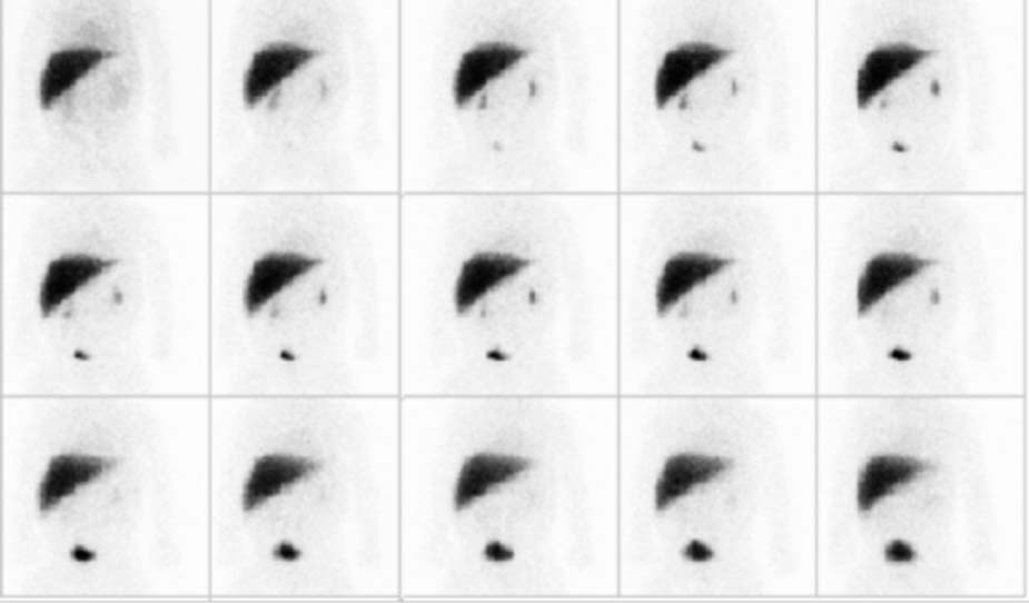
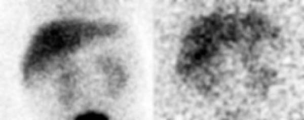
CASE 161 A hepatobiliary scan is requested to help differentiate between biliary atresia and neonatal hepatitis in a 2-week-old infant with extreme lethargy, poor feeding, and a high direct bilirubin level. Sepsis and metabolic disorders have been ruled out. Fig. 161.1 Fig. 161.2 • 99mTc-disofenin is administered intravenously at a dose of 0.05 mCi/kg at the beginning of the acquisition. The minimum dose is 0.25 mCi, and the maximum dose is 3.0 mCi. • Use a high-resolution or ultra-high-resolution, low-energy, parallel-hole collimator. • Energy window is 20% centered at 140 keV. • Imaging time for the initial series is 0.5 minute per frame for 60 minutes. Additional images are obtained at 4 and 24 hours. • Pre-treatment with phenobarbital is administered at 2.5 mg/kg twice a day for 3 to 5 days before hepatobiliary scintigraphy. Images displayed as 5-minute frames after the injection of 99mTc-disofenin (Fig. 161.1) demonstrate prompt hepatic uptake of tracer. The high early liver-to-background ratio and the clear definition of the liver boundaries indicate that there is good hepatic extraction of 99mTc-disofenin, although the prominent renal excretion suggests a mild degree of hepatic dysfunction. Tracer is not identified in the small intestine. Images at 4 and 24 hours (Fig. 161.2) show no evidence of tracer in the bowel. • Biliary atresia • Severe hepatocellular disease • Complete common duct obstruction • Intrahepatic cholestasis
Clinical Presentation
Technique
Image Interpretation
Differential Diagnosis
Related posts:


Stay updated, free articles. Join our Telegram channel

Full access? Get Clinical Tree


