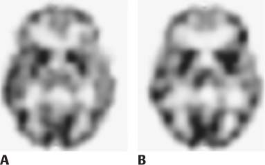
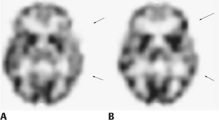
CASE 162 A 6-year-old girl with a 4-year history of partial complex seizures that proved refractory to medical treatment is being considered for surgical cortical resection of an epileptogenic focus pending its localization. Results of CT and MRI of the brain are within normal limits. Electroencephalography is nonlocalizing. A SPECT study with 99mTc-bicisate is obtained. Fig. 162.1 Fig. 162.2 • 99mTc-bicisate is administered at a dose of 0.3 mCi/kg of body weight. The minimum dose is 5 mCi; the maximum dose is 20 mCi. • Use an ultra-high-resolution collimator. • Energy window is 20% centered at 140 keV. • SPECT acquisition on a 128 × 128 matrix with a triple-detector system takes approximately 20 minutes. A usual technique includes 40 stops over 360 degrees per detector, 20 to 30 seconds per stop. • The ictal study is performed in an inpatient setting, preferably in an epilepsy monitoring unit. Continuous electroencephalography (EEG) and video telemetry are essential to ensure ictal injection of tracer. Venous access is established before any seizure activity so that radiotracer can be quickly injected at the onset of a seizure. Interictal perfusion brain SPECT (Fig. 162.1A) demonstrates relatively low regional blood flow to the left temporal and posterior frontal regions. Ictal brain SPECT (Fig. 162.1B) reveals a well-defined increase in blood flow to the left temporal and posterior frontal regions. These correspond to the areas of decreased perfusion on the interictal study. Arrows indicate the location of the finding on the interictal (Fig. 162.2A) and ictal (Fig. 162.2B) images. • Epilepsy • Encephalitis
Clinical Presentation
Technique
Image Interpretation
Differential Diagnosis
Stay updated, free articles. Join our Telegram channel

Full access? Get Clinical Tree


