Case 17
 Clinical Presentation
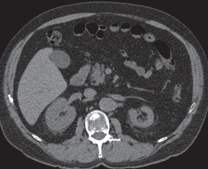
A 62-year-old man with an incidental finding on computed tomography.
 Imaging Findings
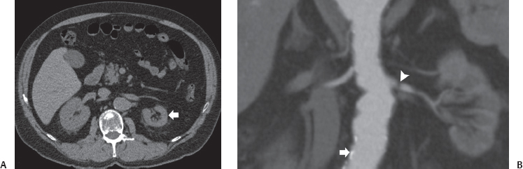
(A) Noncontrast computed tomography (CT) image at the level of the kidneys shows that the left kidney (arrow) is significantly smaller than the right. The outline of the unilateral small kidney is smooth. No dilatation of the collecting system is seen. (B) CT angiographic image at the level of the origin of renal arteries shows atherosclerotic changes in the aorta (arrow). The left renal artery has a tight stenosis (arrowhead).
 Differential Diagnosis
• Small, smooth kidney due to renal vascular disease: It may be due to renal artery stenosis, thrombosis, or embolism or to renal vein thrombosis. The small kidney retains its smooth outline. The collecting system is of normal size and not dilated.
• Postobstructive atrophy:
Stay updated, free articles. Join our Telegram channel

Full access? Get Clinical Tree


